Quick Answer for H. Pylori Recovery Diet: An effective H. pylori post-antibiotic recovery diet prioritizes low-acid, low-fat, easily digestible meals (GSI score 70–100) for 6 to 12 weeks. Focus on soluble beta-glucan starches, lean proteins, and sulforaphane-rich cooked vegetables to rebuild the gastric mucin layer while avoiding acidic fermented foods, caffeine, and heavy lipids that trigger pepsin activation.
Not a treatment protocol
This guide has not been reviewed by a licensed clinician. Specific figures are illustrative, and no food score is a prescription. Talk to your gastroenterologist before changing your diet or medication. See our editorial standards.
Completing a 10-to-14-day course of triple or quadruple therapy for Helicobacter pylori (H. pylori) is one of the most grueling medical interventions in outpatient gastroenterology. Patients endure heavy systemic loads of clarithromycin, amoxicillin, metronidazole, or tetracycline, alongside bismuth subsalicylate and high-dose Proton Pump Inhibitors (PPIs). When the final pill is swallowed, there is a natural expectation that gastric pain, early satiety, burning, and nausea will immediately cease.
In clinical practice, the opposite frequently occurs. Patients enter what gastroenterologists recognize as the post-antibiotic paradox: the pathogen has been eradicated, yet the stomach feels more reactive, raw, and unstable than it did prior to diagnosis.
Understanding why this happens requires looking at the microscopic architecture of the stomach after H. pylori colonization. H. pylori survives in the acidic gastric lumen by secreting urease, an enzyme that converts urea into ammonia and carbon dioxide. While this neutralizes acid immediately surrounding the bacterium, the ammonia—combined with bacterial cytotoxins like VacA (vacuolating cytotoxin A) and CagA (cytotoxin-associated gene A)—systematically degrades the host’s protective mucous layer. It disrupts intercellular tight junctions, depletes mucosal prostaglandins, and triggers a dense infiltration of neutrophils and mononuclear inflammatory cells into the lamina propria.
Antibiotics clear the bacterial infection, stopping the ongoing production of ammonia and bacterial virulence factors. However, antibiotics do nothing to reconstruct the eroded mucin-bicarbonate barrier. Furthermore, broad-spectrum antimicrobial therapy decimates the commensal microbiome of both the upper and lower gastrointestinal tract, altering motility and visceral sensitivity.
When a patient finishes therapy and immediately resumes a standard diet—or worse, aggressively introduces acidic fermented foods in an attempt to "fix their microbiome"—they expose denuded, hyper-sensitized gastric epithelium directly to hydrochloric acid ($\text{HCl}$), active pepsin, and bile salts.
Recovery from H. pylori requires treating the post-eradication stomach as an acute internal wound. This guide outlines the clinical physiology of post-eradication gastritis and provides a structured, three-phase nutritional protocol anchored by the Gastric Safety Index (GSI) to rebuild your stomach lining safely.
The Pathophysiology of the Post-Eradication Stomach
To navigate dietary recovery, you must understand the three overlapping physiological challenges occurring inside your stomach the day after antibiotic therapy ends.
1. Mucin-Bicarbonate Barrier Depletion and Pepsin Activation
A healthy stomach protects itself from digesting its own tissue via a dual-layer mucus barrier secreted by surface foveolar cells. The inner layer is dense, firmly adherent, and rich in phospholipids and bicarbonate ($\text{HCO}_3^-$), which maintains a neutral pH ($\approx 7.0$) directly against the epithelial cell membranes, even when the gastric lumen pH drops below $2.0$.
During H. pylori infection, bacterial mucolytic enzymes degrade the polymeric structure of mucin glycoproteins (primarily MUC5AC and MUC6). After eradication, it takes weeks for goblet and foveolar cells to upregulate mucin synthesis and restore normal layer thickness. Until this layer is restored, luminal acid diffuses backward into the tissue (acid back-diffusion).
Crucially, when luminal pH drops below $4.0$, chief cells' inactive zymogen, pepsinogen, cleaves into active pepsin. Pepsin is a proteolytic enzyme designed to digest dietary collagen and muscle fibers. When the mucus barrier is thin, pepsin digests the exposed mucosal proteins of your own stomach wall, causing chemical gastritis, micro-erosions, and epigastric burning.
2. Rebound Acid Hypersecretion (RAHS)
Standard H. pylori eradication regimens require twice-daily high-dose PPI therapy (e.g., esomeprazole or pantoprazole $40\text{ mg}$ BID) to keep intragastric pH above $5.0$. This elevated pH forces H. pylori into a replicative state, making it susceptible to antibiotics like amoxicillin and clarithromycin.
However, sustained suppression of gastric acid removes the natural negative feedback loop on gastrin production. In response to high pH, antral G-cells secrete large amounts of the hormone gastrin into the bloodstream (hypergastrinemia). Gastrin stimulates Enterochromaffin-like (ECL) cells to hypertrophy and proliferate.
When you abruptly stop taking PPIs at the end of your antibiotic course—or when you must stop them for 14 days prior to your confirmation urea breath test or stool antigen test—those expanded ECL cell populations release massive amounts of histamine. This drives parietal cells into overdrive, resulting in Rebound Acid Hypersecretion (RAHS). Your stomach produces significantly more acid than it did at baseline, pouring high-titratable acid directly onto an unhealed mucosal surface.
3. Antibiotic-Induced Dysbiosis and Motility Disturbance
Macrolide and nitroimidazole antibiotics do not discriminate between H. pylori in the gastric antrum and beneficial Bifidobacterium, Lactobacillus, and butyrate-producing Clostridia species in the small and large intestines. Furthermore, macrolides like clarithromycin act as motilin receptor agonists, forcefully accelerating upper GI contractions during treatment, which often leaves the enteric nervous system dysregulated afterward.
The result is delayed gastric emptying (functional gastroparesis), visceral hypersensitivity, and Small Intestinal Bacterial Overgrowth (SIBO) vulnerability—especially while gastric acid remains suppressed.
The Three Phases of Mucosal Healing
Because histological recovery occurs in distinct biological stages, a static "gastritis diet" fails. Eating a diet that is too restrictive for four months leads to micronutrient deficiencies and unintentional weight loss; advancing your diet too early triggers acute inflammatory flares that reset your healing timeline.
At Gutrack, we categorize post-H. pylori nutritional rehabilitation into three clinical phases based on mucosal histology and meal Gastric Safety Index (GSI) scores.
Phase 1: Acute Post-Eradication & Barrier Protection (Days 1–14)
- Histological Reality: The bacteria are dead or dying, but polymorphonuclear neutrophil infiltration is still clearing. The mucus layer is patchy, intercellular tight junctions (claudins and occludins) are leaky, and the patient is often tapering off PPIs or entering the window for eradication testing.
- Clinical Goal: Minimize gastric residence time, prevent pepsin activation, and provide physical colloidal coating to the mucosa.
- Target Meal GSI Score: 80–100 (Strict Safe Zone).
- Dietary Protocol:
- All vegetables must be thoroughly cooked, peeled, and deseeded. Raw insoluble fiber acts as a mechanical abrasive against hyperemic (red, engorged) mucosa.
- Dietary fat must be restricted to $<10\text{ grams}$ per meal. Lipids entering the duodenum stimulate I-cells to release cholecystokinin (CCK), a hormone that potently slows gastric emptying and induces transient lower esophageal sphincter relaxations (TLESRs). Slower emptying means food, acid, and pepsin sit against raw stomach tissue for hours.
- Meals must be small in volume ($250\text{–}350\text{ mL}$), consumed every 3 hours to utilize food proteins as a natural pH buffer without distending the gastric fundus.
Phase 2: Epithelial Regeneration & Tight Junction Repair (Weeks 3–6)
- Histological Reality: Surface epithelial cells have turned over several times. Acute neutrophil infiltration has resolved, though chronic mononuclear infiltrates (lymphocytes) remain in the lamina propria. If the patient stopped PPIs for their 4-week confirmation test, RAHS typically peaks between weeks 2 and 4 post-cessation before gradually normalizing.
- Clinical Goal: Supply targeted amino acids, trace minerals, and phytonutrients to synthesize mucin glycoproteins and seal tight junctions, while managing acid rebound.
- Target Meal GSI Score: 70–85 (Safe Zone).
- Dietary Protocol:
- Introduce gentle, non-acidic prebiotic fibers and lightly steamed cruciferous sprouts (specifically for sulforaphane).
- Gradually increase healthy monounsaturated and omega-3 fats (e.g., extra virgin olive oil, baked salmon) to $12\text{–}15\text{ grams}$ per meal to support prostaglandin $E_2$ ($\text{PGE}_2$) synthesis, which regulates mucosal blood flow and bicarbonate secretion.
- Maintain a strict ban on caffeine, alcohol, capsaicin, and organic acids (citric, malic, and acetic acid).
Phase 3: Microbiome Restoration & Functional Expansion (Months 2–4)
- Histological Reality: Mucin thickness has returned to baseline, and ECL cell hyperplasia from prior PPI use is subsiding. Gastric acid output is normalizing, restoring the stomach's natural chemical barrier against ingested pathogens.
- Clinical Goal: Rebuild microbial diversity in the lower gut, re-establish normal enzymatic secretion, and test moderate-risk foods systematically.
- Target Meal GSI Score: 60–80 (Safe to Upper Caution Zone).
- Dietary Protocol:
- Begin controlled reintroduction of raw soluble fibers (e.g., peeled apples, ripe berries), diverse plant polyphenols, and small, diluted quantities of fermented foods with meals (never on an empty stomach).
- Transition from 5–6 small grazing meals back to 3 structured meals and 1 snack per day to allow the Migrating Motor Complex (MMC)—the gut's inter-digestive housekeeping wave—to sweep residual bacteria out of the small intestine every 90 to 120 minutes during fasting states.
Nutritional Building Blocks for Mucosal Repair
Rebuilding the stomach lining is an energy-intensive anabolic process. Rather than viewing your diet merely as a list of triggers to avoid, you must view your safe meals as delivery vehicles for specific biochemical substrates.
1. Sulforaphane and Residual Bacterial Suppression
Even after successful antibiotic therapy, trace pockets of coccoid (dormant) H. pylori or localized oxidative stress can persist. Sulforaphane, an isothiocyanate derived from the hydrolysis of glucoraphanin by the enzyme myrosinase, is one of the most heavily researched phytonutrients in gastroenterology.
Clinical trials (such as those conducted by Yanaka et al., published in Cancer Prevention Research) demonstrate that sulforaphane exhibits potent bactericidal activity against H. pylori—including clarithromycin-resistant strains—and activates the Nrf2 pathway in human gastric mucosal cells. Nrf2 upregulates phase II antioxidant enzymes (like heme oxygenase-1 and glutathione S-transferase), neutralizing the reactive oxygen species (ROS) that cause post-infectious mucosal atrophy.
- Clinical Preparation Rule: Broccoli sprouts contain up to 50 times more glucoraphanin than mature broccoli heads. However, eating raw sprouts in Phase 1 can cause gas and mechanical irritation, while boiling them destroys the myrosinase enzyme required to make sulforaphane bioavailable. The solution: Steam broccoli florets or sprouts lightly at $70^\circ\text{C}$ ($158^\circ\text{F}$) for no more than 3 minutes, or blend raw sprouts into a smooth, warm (not boiling) potato or squash puree.
2. L-Glutamine and Mucosal Enterocyte Fuel
L-glutamine is a conditionally essential amino acid that serves as the primary metabolic fuel for rapidly dividing gastrointestinal epithelial cells. In the stomach and duodenum, glutamine stimulates the synthesis of hexosamines, which are the structural building blocks of mucin glycoproteins. Furthermore, glutamine upregulates the expression of tight junction proteins (occludin and ZO-1), directly reducing paracellular permeability ("leaky gut" caused by antibiotics).
- Dietary Sources: Poached chicken breast, turkey tenderloin, cod, halibut, egg whites, and homemade bone broth that has been chilled and completely skimmed of its solid surface lipid layer.
3. Beta-Glucans and Colloidal Mucoadhesion
While the stomach regenerates its endogenous mucus layer, you can provide an exogenous physical barrier using soluble, viscous polysaccharides—specifically beta-glucans and mucilages. When cooked with water, beta-glucans form a soothing, gelatinous hydrocolloid matrix. This matrix adheres temporarily to the gastric mucosa, creating a physical buffer that slows the diffusion of $\text{HCl}$ and pepsin toward the vulnerable epithelial surface.
- Dietary Sources: Rolled or steel-cut oats cooked to a soft porridge consistency, barley congee, peeled and boiled white potatoes (cooled slightly to increase resistant starch for the colon), and marshmallow root or slippery elm infusions taken between meals.
4. Zinc-L-Carnosine and Prostaglandin Synthesis
Zinc is an essential cofactor for DNA polymerase and cellular proliferation during wound healing. In clinical practice, the chelated compound Zinc-L-Carnosine (polaprezinc) is widely utilized alongside dietary zinc. Unlike standard zinc sulfate, which dissociates rapidly in stomach acid and can cause severe nausea, Zinc-L-Carnosine adheres selectively to ulcerated or eroded lesions via electrostatic attraction to exposed tissue proteins, releasing zinc and L-carnosine directly into the damaged site to stimulate mucus production and quench free radicals.
- Dietary Sources: Well-cooked lean beef tenderloin (Phase 2/3), pumpkin seed butter (smooth, unsalted), and white fish.
The Gastric Safety Index (GSI) Post-H. Pylori Food Matrix
When recovering from H. pylori, patients frequently fall into the trap of binary food lists ("good foods" vs. "bad foods"). However, gastric physiology does not operate in black and white; it operates on thresholds of acidity, osmolarity, lipid load, and mechanical texture.
A food that is theoretically healthy—such as a raw kale salad with lemon-olive oil dressing and fermented sauerkraut—can trigger a debilitating 48-hour gastritis flare in a post-antibiotic stomach. Why? Because the raw cellulose scrapes the mucosa, the citric and lactic acids activate pepsinogen into pepsin immediately upon contact, and the lipid load delays gastric emptying.
To eliminate guesswork, Gutrack evaluates meals using the Gastric Safety Index (GSI), a clinical scoring algorithm ranging from 0 to 100. The GSI weighs four primary physiological variables:
- Titratable Acidity & pH: Potential to drop intragastric pH below the $4.0$ pepsin activation threshold.
- Lipid Density & CCK Load: Potential to delay gastric emptying and relax the lower esophageal sphincter.
- Mechanical & Osmotic Irritancy: Fiber insolubility, particulate hardness, spice capsaicinoids, and hyperosmolar sugar/salt concentrations.
- Mucosal Buffering Capacity: Presence of soluble hydrocolloids and non-irritating protein buffers.
- 70–100 (Safe Zone): Ideal for Phases 1 and 2. Gentle on the stomach lining, promotes predictable gastric emptying, and buffers acid.
- 40–69 (Caution Zone): Appropriate for Phase 3 testing. Requires intact mucosal barrier function and controlled portion sizes.
- 0–39 (High Risk Zone): Strictly contraindicated during recovery. Triggers immediate pepsin activation, bile reflux, or neurogenic inflammation.
| Meal / Food Item | GSI Score (0–100) | Clinical Zone | Physiological & Biochemical Mechanism in Post-H. Pylori Mucosa |
|---|---|---|---|
| Steamed Cod with Pureed Butternut Squash & White Rice | 94 / 100 | Safe (Phase 1–3) | Ultra-low lipid content (<2g fat) prevents CCK-mediated delayed emptying. Digestible marine proteins buffer luminal HCl without requiring excessive pepsinogen secretion. Soluble squash pectin coats foveolar cells. |
| Warm Rolled Oat Porridge with Mashed Ripe Banana & Pea Protein | 91 / 100 | Safe (Phase 1–3) | Aqueous beta-glucans form a protective colloidal film over denuded mucosa. Ripe banana provides natural leukocyanidin (a flavonoid that increases mucus thickness) at a safe pH (~5.0). |
| Poached Chicken Breast in Skimmed Bone Broth with Boiled Carrots | 88 / 100 | Safe (Phase 1–3) | High bioavailable L-glutamine and glycine directly fuel enterocyte tight junction synthesis (ZO-1/occludin). Boiling breaks down carrot hemicellulose, eliminating mechanical friction. |
| Lightly Steamed Broccoli Sprouts over Mashed White Potatoes (1 tsp Olive Oil) | 82 / 100 | Safe (Phase 2–3) | Delivers active sulforaphane to upregulate Nrf2 antioxidant pathways and suppress residual bacterial urease. Potato starch buffers gastric acid; minimal olive oil supports PGE2 synthesis without slowing transit. |
| Baked Wild Salmon with Steamed Asparagus Tips & Quinoa | 74 / 100 | Safe (Phase 2–3) | Rich in EPA/DHA omega-3 fatty acids, resolving lymphocytic mucosal inflammation. Slightly lower GSI than white fish due to higher lipid load (~12g fat), which modestly slows antral peristalsis. |
| Sourdough Toast with 1 Whole Poached Egg & Mashed Avocado (1/4 fruit) | 64 / 100 | Caution (Phase 3) | Fermented sourdough reduces phytic acid and fructans, making it easier to digest than yeast bread, but combined fats from egg yolk and avocado (~14g) stimulate moderate CCK release. Test in smaller portions. |
| Plain Low-Fat Greek Yogurt with Blueberries & Raw Honey | 52 / 100 | Caution (Phase 3) | Contains lactic acid (pH ~4.3–4.5), which sits near the pepsin activation threshold. Raw honey is hyperosmolar, which can draw fluid into the gastric lumen and cause transient cramping if the epithelium is still thin. |
| Raw Kale & Apple Salad with Lemon Vinaigrette & Walnuts | 34 / 100 | High Risk (Avoid) | Dense insoluble cellulose requires vigorous mechanical trituration in the antrum, scraping inflamed tissue. Lemon juice (citric acid, pH ~2.2) instantly activates pepsin directly on food contact. |
| Spicy Kimchi or Raw Milk Kefir (Taken on an Empty Stomach) | 18 / 100 | High Risk (Avoid) | High titratable organic acids (lactic/acetic, pH <3.8) burn unprotected subepithelial nerve endings. Capsaicin in kimchi binds to TRPV1 pain receptors, triggering acute neurogenic inflammation and burning. |
| Espresso or Dark Roast Coffee with Pastries / Croissants | 08 / 100 | High Risk (Avoid) | Chlorogenic acids and caffeine potently stimulate parietal cell H+/K+ ATPase proton pumps, worsening post-PPI acid rebound. High saturated pastry fats halt gastric emptying and drop LES pressure. |
Why Food Logging Without GSI Scores Fails
During H. pylori recovery, patients are often told by their clinicians to "keep a food diary." Typically, a patient writes down notes in a standard notebook or basic tracking app: Breakfast: Yogurt and berries. Lunch: Chicken soup. Dinner: Salmon and salad. Symptoms: Severe burning at 4:00 PM.
Because gastric emptying takes 2 to 4 hours—and delayed mucosal inflammation can peak 6 to 12 hours after a trigger—manual food diaries quickly become overwhelming. Everything starts to feel like a trigger. Patients develop food fear, stripping their diet down to plain white rice and water, which starves the stomach lining of the exact proteins and micronutrients required for tissue repair.
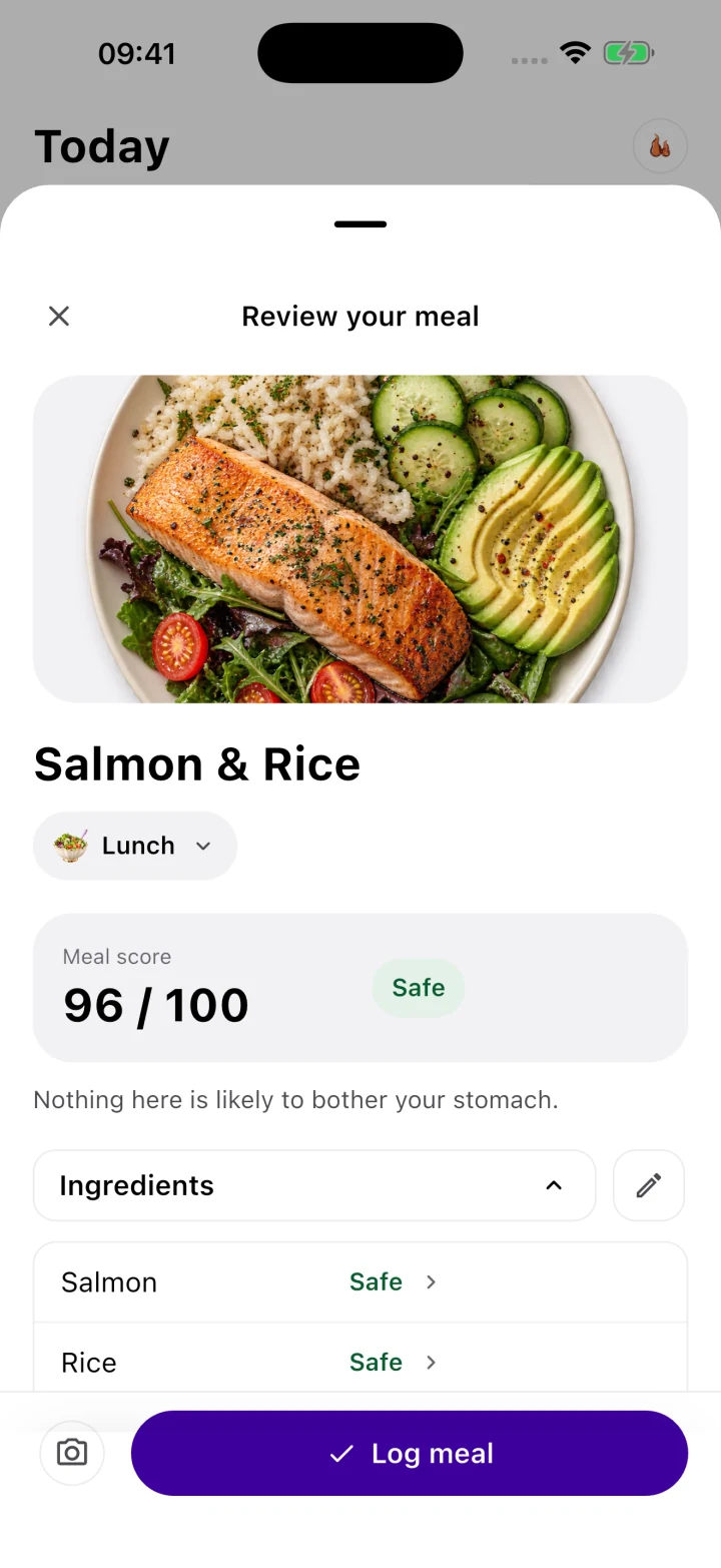
This is why we built the Gastric Safety Index into the Gutrack iOS app. Instead of guessing whether a meal is safe, you can scan or log your meal in Gutrack to receive an instant 0–100 GSI score before you take a bite. The algorithm flags hidden physiological triggers—such as excessive lipid load, high titratable acidity, or abrasive fiber combinations—and correlates your daily GSI trends directly against your symptom curve.
Restoring the Gastric Microbiome Safely: The Fermented Food Trap
One of the most common clinical errors we see in post-H. pylori patients is the premature aggressive consumption of fermented foods. Driven by well-intentioned advice that "antibiotics destroy your gut bacteria, so you need probiotics," patients finish their quadruple therapy and immediately start drinking kombucha, eating sauerkraut, or taking shots of apple cider vinegar and kefir.
Within 48 hours, they are frequently in severe epigastric pain, convinced their H. pylori infection has returned.
The Biochemistry of Premature Fermentation
While fermented foods are beneficial for a healthy, intact gastrointestinal tract, they present three distinct biochemical hazards to a post-antibiotic stomach:
- High Titratable Acidity: Fermentation works because bacteria convert sugars into organic acids—primarily lactic acid and acetic acid—dropping the pH of the food to between $3.2$ and $4.2$ to prevent spoilage. When you pour a liquid with a pH of $3.5$ into a stomach lacking its mucin barrier, it immediately converts luminal pepsinogen into active pepsin and chemically irritates exposed mucosal nerve fibers.
- Biogenic Amines and Histamine: Fermented foods (especially aged cheeses, kimchi, sauerkraut, and kombucha) contain high levels of histamine and tyramine produced by microbial decarboxylation of amino acids. In the stomach, histamine binds directly to H2 receptors on parietal cells, stimulating hydrochloric acid secretion. If you are already experiencing post-PPI Rebound Acid Hypersecretion, dietary histamine pours fuel on the fire.
- D-Lactic Acid Production: Certain Lactobacillus species found in commercial ferments produce D-lactic acid, which mammals metabolize slowly. In a post-antibiotic gut with altered motility, rapid fermentation of carbs by these strains can cause severe upper abdominal distension and gas pressure against the lower esophageal sphincter.
The Clinical Protocol for Microbiome Rebuilding
Instead of using acidic fermented foods during Phases 1 and 2, rebuild your microbiome using a two-step medical nutrition approach:
Step 1: Encapsulated Targeted Probiotics (Weeks 1–8)
Use purified, lyophilized probiotic supplements enclosed in delayed-release capsules. These bypass the raw gastric mucosa without introducing organic acids or histamine. Look for strains backed by randomized controlled trials for H. pylori recovery:
- Saccharomyces boulardii (specifically strain CNCM I-745): A non-pathogenic probiotic yeast that is naturally resistant to antibiotics. It binds bacterial toxins, stimulates secretory IgA (sIgA) to protect the mucosal surface, and prevents opportunistic Clostridioides difficile overgrowth.
- Lactobacillus reuteri (DSM 17648 / ATCC 55730): Demonstrated in clinical studies to co-aggregate with H. pylori in the stomach and suppress post-eradication inflammatory cytokines (such as IL-8 and TNF-$\alpha$) in the gastric mucosa.
- Bifidobacterium longum and Lactobacillus rhamnosus GG: Essential for restoring colonic butyrate production and resolving antibiotic-associated bowel irregularity.
Step 2: Soluble Prebiotic Fibers Before Ferments (Weeks 4–12)
Before throwing live fermented foods into your gut, feed your surviving endogenous bacteria with low-fermentation, non-acidic soluble fibers. Cooked and cooled white potatoes (rich in Type 3 resistant starch), steamed carrot puree, oat beta-glucans, and partially hydrolyzed guar gum (PHGG) pass harmlessly through the stomach with a neutral pH (GSI > 85) and ferment slowly in the colon to produce butyrate—a short-chain fatty acid that regulates systemic mucosal immunity.
Only when you have maintained a symptom-free baseline for at least 3 consecutive weeks in Phase 3 should you test fermented foods—starting with a single teaspoon of plain yogurt or diluted kefir folded into a warm bowl of oatmeal (buffering the pH), never on an empty stomach.
Managing the Post-Antibiotic PPI Taper and Testing Window
A critical medical reality of H. pylori treatment is the confirmation of eradication test. Because global antibiotic resistance rates (particularly to clarithromycin and levofloxacin) now exceed 20–30% in many regions, gastroenterological guidelines (such as the Maastricht VI/Florence Consensus Report and American College of Gastroenterology guidelines) mandate that every patient undergo testing—via a Urea Breath Test (UBT), fecal antigen test, or upper endoscopy—to prove the bacteria are gone.
The 14-Day PPI Washout Challenge
To prevent a false-negative result, you must completely stop taking Proton Pump Inhibitors for at least 14 days (and stop bismuth and antibiotics for at least 28 days) prior to the test. PPIs suppress bacterial density and urease activity; testing while on a PPI frequently makes an active infection look cured.
However, stopping a high-dose PPI cold turkey right after antibiotic therapy triggers the exact Rebound Acid Hypersecretion (RAHS) described earlier. Patients experience a sudden surge in acid burning 3 to 7 days after stopping their PPI and panic, assuming their treatment failed.
How to Survive the Testing Washout Safely:
- Strict Phase 1 GSI Adherence (Score > 80): During the 14-day PPI washout window, your diet must act as your primary acid buffer. Eat small, protein-and-starch meals every 3 hours (e.g., poached egg whites with white rice, steamed cod, oatmeal). Dietary proteins bind free $\text{H}^+$ ions in the gastric lumen, temporarily raising intragastric pH without interfering with your upcoming breath or stool test.
- Utilize Allowed Bridge Medications (With Physician Approval): While PPIs must be stopped 14 days before testing, H2-receptor antagonists (such as famotidine) typically only need to be stopped 24 to 48 hours before a breath test (confirm your specific lab’s protocol with your gastroenterologist), and standard calcium/magnesium antacids or sodium alginate rafts can often be used up until the day before testing to physically block acid reflux without altering bacterial counts.
- Positional Mechanics: Never lie flat within 3 hours of your final evening meal, and elevate the head of your bed by 6 inches. Because RAHS increases nocturnal acid volume, gravity is essential to prevent nocturnal acid contact with the lower esophageal sphincter and gastric cardia.
Tracking Post-Treatment Symptoms with Gutrack: Healing vs. Recurrence
During the 8 to 16 weeks required for full mucosal remodeling, healing is never strictly linear. You will experience good days followed by sudden symptomatic dips. For the patient, the most stressful question is always: "Did I just eat something that irritated my healing stomach lining, or did the antibiotics fail and the H. pylori is still there?"
Answering this question requires looking at objective data over a 14-to-28-day timeline rather than reacting emotionally to a single bad afternoon.
Distinguishing Post-Infectious Gastritis from True Failure
In clinical practice, the symptom patterns of mucosal healing look remarkably different from active bacterial failure when plotted alongside dietary GSI scores:
- Pattern A: Mucosal Healing with Dietary/RAHS Flares (Bacteria Eradicated)
- Baseline symptoms gradually trend downward week over week when meal GSI scores are kept above 75.
- Pain or burning occurs predictably 1 to 3 hours after a clear physiological trigger (e.g., a restaurant meal with hidden seed oils/butter dropping the GSI to 45, a stressful skip in meals allowing unbuffered acid to pool, or day 5 of PPI cessation).
- Returning to Phase 1 safe foods for 48 hours reliably calms the flare.
- Pattern B: Persistent Infection or Refractory Pathology (Requires Medical Re-evaluation)
- Epigastric pain, waking at 2:00 AM with gnawing hunger-like pain (classic duodenal/antral hyperacidity), and nausea remain constant at a 6/10 or higher for weeks, even when your 14-day average GSI score is consistently above 85.
- Progressive unintentional weight loss, dysphagia (difficulty swallowing), persistent vomiting, or dark, tarry stools (melena).
Turning Subjective Symptoms into Clinical Evidence
When you return to your gastroenterologist for your post-treatment follow-up, saying "My stomach still hurts a lot, I think the antibiotics didn't work" leaves the physician with very little actionable data. They cannot tell if you are experiencing expected post-PPI rebound, dietary irritation from hidden triggers, delayed gastric emptying, or true treatment failure.
With Gutrack, you eliminate this communication gap. By scanning your daily meals to log their 0–100 Gastric Safety Index scores alongside timestamped symptom severity (epigastric burning, early satiety, bloating, nausea) and medication tapers, you build a longitudinal dataset.
Before your appointment, you can export a 14-day Objective Clinical PDF Report directly from the app. This report gives your gastroenterologist a clean, one-page visual correlation between your dietary mucosal load (GSI), your PPI washout timeline, and your symptom curve. Instead of guessing, your doctor can immediately see whether your stomach is responding appropriately to a low-acid, low-lipid protocol or if targeted prokinetics, sucralfate mucosal coating, or second-line susceptibility testing is warranted.
Sample 3-Day Phase 1 Post-Antibiotic Recovery Menu (Average GSI: 89)
To help you implement this protocol immediately upon finishing your antibiotic therapy, follow this structured 3-day Phase 1 template. Notice that meals are divided into 5 smaller feedings to prevent antral distension while keeping protein buffers present in the stomach throughout the day.
Day 1
- 8:00 AM (Breakfast – GSI 91): 1 cup cooked rolled oats made with water or almond milk (fortified, carrageenan-free), topped with $\frac{1}{2}$ mashed ripe banana and 1 scoop unflavored pea or rice protein isolate.
- 11:00 AM (Mid-Morning Buffer – GSI 94): 1 cup warm, fat-skimmed chicken bone broth with $\frac{1}{2}$ cup soft-boiled white rice and 2 oz shredded poached chicken breast.
- 2:00 PM (Lunch – GSI 92): 4 oz steamed cod or tilapia seasoned with a pinch of sea salt and dried dill; 1 cup pureed butternut squash; $\frac{1}{2}$ cup peeled, boiled zucchini slices.
- 5:00 PM (Afternoon Buffer – GSI 88): 2 scrambled egg whites (cooked in a ceramic non-stick pan with a mist of olive oil) on 1 slice of toasted white sourdough or gluten-free rice bread.
- 7:30 PM (Dinner – GSI 89): 4 oz baked turkey breast tenderloin; 1 cup mashed peeled white potatoes (whipped with warm potato cooking water and $\frac{1}{2}$ tsp extra virgin olive oil); $\frac{1}{2}$ cup steamed carrot coins.
- Between Meals: Sip warm chamomile tea or slippery elm bark infusion ($30\text{ minutes}$ before meals or $1\text{ hour}$ after).
Day 2
- 8:00 AM (Breakfast – GSI 93): Warm cream of rice or rice congee cooked with 2 liquid egg whites stirred in at the end for protein, topped with $\frac{1}{4}$ cup steamed, peeled pear puree.
- 11:00 AM (Mid-Morning Buffer – GSI 87): Smoothie made with 1 cup oat milk, $\frac{1}{2}$ ripe papaya (seeded and peeled—papaya contains gentle enzymes and supports mucin synthesis), 1 scoop collagen peptides or hemp protein, and $\frac{1}{4}$ cup cooked oats (blended at room temperature, never ice-cold).
- 2:00 PM (Lunch – GSI 90): 4 oz poached chicken breast over 1 cup white jasmine rice and $\frac{1}{2}$ cup lightly steamed spinach (chopped finely to reduce fiber length).
- 5:00 PM (Afternoon Buffer – GSI 92): 2 plain rice cakes topped with 2 oz flaked canned tuna (packed in water, drained) mixed with 1 tbsp pureed avocado.
- 7:30 PM (Dinner – GSI 84): 4 oz baked halibut; 1 cup mashed sweet potato; $\frac{1}{4}$ cup lightly steamed broccoli sprouts (steamed for 2–3 minutes to preserve myrosinase/sulforaphane).
Day 3
- 8:00 AM (Breakfast – GSI 90): 1 cup oatmeal porridge cooked with 1 tsp smooth almond butter stirred in thoroughly (diluting the lipid load across the aqueous starch matrix) and a dash of Ceylon cinnamon.
- **11:00 AM (Mid-Morning Buffer – GSI
Self-tracking tool, not clinical diagnosis
Gutrack provides evidence-based Gastric Safety Index estimates for daily self-management and diet optimization. It does not replace medical consultation, endoscopy, or treatment prescribed by a qualified gastroenterologist.
Frequently asked questions
How long does it take for the stomach lining to heal after H. pylori eradication?
Superficial epithelial cells regenerate every 3 to 7 days, but deep mucosal remodeling and the resolution of chronic inflammatory infiltrates (lymphocytes and plasma cells) require 8 to 16 weeks after successful antibiotic therapy. Functional symptom resolution typically lags behind histological clearance by 1 to 3 months.
Why do I still have gastritis symptoms after finishing H. pylori antibiotics?
Antibiotics kill the bacteria, not the inflammation. Residual symptoms occur because the protective mucin-bicarbonate barrier remains depleted, exposing subepithelial nerve endings to gastric acid and pepsin, while high-dose Proton Pump Inhibitor (PPI) withdrawal often triggers temporary rebound acid hypersecretion.
What foods help rebuild stomach mucosa after H. pylori?
Foods rich in soluble beta-glucans (rolled oats), L-glutamine (poached poultry, bone broth skimmed of fat), and sulforaphane (lightly steamed broccoli sprouts) actively support mucosal repair. These foods provide a physical colloidal buffer against gastric acid while supplying amino acids for tight junction synthesis.
Should I take probiotics during or after H. pylori treatment?
Clinical trials show that targeted probiotic strains like Saccharomyces boulardii CNCM I-745 and Lactobacillus reuteri DSM 17648 reduce antibiotic-associated diarrhea and improve eradication rates. However, patients should use encapsulated, non-acidic supplements rather than dietary fermented foods (like kefir or kimchi), which contain high titratable acidity that irritates raw gastric tissue.
During post-antibiotic recovery, targeted nutrition accelerates mucosal healing. Our evidence review of bone broth, gelatin, and L-glutamine separates the structural amino acid science from the marketing hype.
Sources & background reading
Background sources for definitions, diagnosis and treatment. Food scores are estimates, not clinical targets. See our editorial standards.
Clinical Evidence & Scientific References
- Chey WD, Leontiadis GI, Howden CW, Moss SF. ACG Clinical Guideline: Treatment of Helicobacter pylori Infection. Am J Gastroenterol. 2017;112(2):212-239.
- Yanaka A, et al. Dietary sulforaphane-rich broccoli sprouts reduce colonization and attenuate gastritis in Helicobacter pylori-infected mice and humans. Cancer Prev Res. 2009;2(4):353-360.
- National Institute of Diabetes and Digestive and Kidney Diseases (NIDDK). Treatment & Mucosal Recovery for H. Pylori Infections. NIH Pub. No. 20-4225.
This article is for educational purposes only and isn't a substitute for professional medical advice, diagnosis, or treatment. Gutrack is a self-tracking tool, not a medical device. If you're experiencing persistent or concerning symptoms, talk to a doctor.