Quick Answer for Gastritis, Anxiety & The Gut-Brain Axis: Gastritis and anxiety interact through the bidirectional gut-brain axis. Chronic mucosal inflammation sensitizes enteric and spinal afferent nerves, causing visceral hypersensitivity where normal digestion feels painful. Simultaneously, sympathetic nervous system activation shunts blood away from the stomach lining, suppresses protective bicarbonate and mucus secretion, and delays gastric emptying.
Every gastroenterologist recognizes the clinical presentation: a patient undergoes an upper endoscopy (esophagogastroduodenoscopy, or EGD) after months of debilitating epigastric burning, early satiety, and postprandial nausea. The patient is convinced they have a bleeding ulcer or advanced gastric pathology. Yet when the histology report returns from pathology, it shows either completely normal gastric mucosa or "mild, chronic inactive gastritis" with no Helicobacter pylori colonization and no macroscopic erosions.
The patient is often told their stomach is fine and that the symptoms are "just stress" or "functional dyspepsia." This dismissal is both frustrating for the patient and physiologically incomplete. The pain is entirely real, often reaching 7 or 8 out of 10 on a clinical visual analog scale. However, the primary driver of the pain has shifted from macroscopic tissue ulceration to a self-sustaining neurobiological feedback loop between the gastric mucosa and the central nervous system.
When epigastric pain strikes after a meal, it immediately triggers the brain’s threat-detection circuitry. For a patient who has suffered for months, a subtle twinge in the upper abdomen sparks acute health anxiety: Did I eat the wrong thing? Is my stomach lining eroding again? Will I be awake all night in pain? Within seconds, the hypothalamus-pituitary-adrenal (HPA) axis and the sympathetic nervous system activate. Heart rate climbs, respiratory rate shallows, and catecholamines (epinephrine and norepinephrine) flood the bloodstream.
To prepare the body for perceived danger, the sympathetic nervous system executes splanchnic vasoconstriction—shunting blood away from the gastrointestinal tract toward skeletal muscle. Within the stomach, this abrupt drop in microcirculatory perfusion starves the mucosal epithelium of oxygen, halts the secretion of protective mucus and bicarbonate, and triggers spasms in the pyloric sphincter. Digestion stalls. Food sits in the antrum, fermenting and stimulating prolonged gastrin release. The physical symptoms worsen, confirming the patient's anxious fear, which drives further sympathetic arousal.
Breaking this cycle requires more than standard acid suppression. It demands an understanding of how mucosal inflammation rewires visceral pain pathways, how autonomic tone alters physical digestion, and how to structure nutrition using objective metrics—like the Gastric Safety Index (GSI)—so the stomach and nervous system can recover simultaneously.
The Neurobiology of Visceral Hypersensitivity
To understand why a stomach with minimal histologic inflammation can hurt as severely as an acute peptic ulcer, we must examine the architecture of gastric nociception (pain signaling).
During an initial bout of acute gastritis—whether triggered by nonsteroidal anti-inflammatory drugs (NSAIDs), H. pylori infection, bile reflux, or severe alcohol exposure—the epithelial barrier is breached. Hydrochloric acid ($HCl$) and activated pepsin penetrate the lamina propria. In response, resident mast cells, eosinophils, and macrophages release a localized storm of inflammatory mediators, including histamine, serotonin (5-HT), bradykinin, prostaglandin $E_2$ ($PGE_2$), and nerve growth factor (NGF).
[Acute Mucosal Injury / Acid Breach]
                 │
                 ▼
[Mast Cell & Eosinophil Degranulation] ──► Releases Histamine, 5-HT, NGF
                 │
                 ▼
[Peripheral Sensitization of TRPV1 / ASIC3] ──► Threshold drops from pH 1.5 to pH 4.0
                 │
                 ▼
[Dorsal Horn "Wind-Up" (Central Sensitization)] ──► Normal meal stretch = 8/10 Pain
                 │
                 ▼
[Amygdala & Insular Cortex Hypervigilance] ──► Sympathetic Surge (Ischemia & Stasis)
Peripheral Sensitization: Lowering the Firing Threshold
Embedded within the muscularis and submucosa of the stomach are primary afferent nerve fibers (primarily unmyelinated C-fibers and thinly myelinated A$\delta$-fibers) that travel via the splanchnic and vagus nerves to the spinal cord and brainstem. These nerve endings are equipped with specialized molecular sensors:
- TRPV1 (Transient Receptor Potential Vanilloid 1): Responds to noxious heat, capsaicin, and extracellular protons (hydrogen ions, $H^+$).
- ASIC3 (Acid-Sensing Ion Channel 3): Detects rapid drops in extracellular pH.
- Mechanoreceptors (Piezo2 and TRP channels): Detect physical stretch and tension within the stomach wall during meal accommodation.
In a healthy stomach, TRPV1 and ASIC3 channels remain quiet unless intraluminal acidity drops to extreme levels ($pH < 1.5$) or the stomach is distended to near-bursting volumes (over 1,200 mL). However, prolonged exposure to mast cell histamine and NGF during a bout of gastritis chemically alters these receptors. The receptors undergo phosphorylation, which dramatically lowers their activation threshold—a phenomenon known as peripheral sensitization.
Once sensitized, a TRPV1 receptor does not wait for a highly corrosive $pH$ of 1.5 to fire; it begins firing rapid pain signals at a mild, physiological $pH$ of 4.0. Similarly, mechanoreceptors that previously ignored the gentle stretch of a 300 mL meal now interpret normal gastric filling as dangerous mechanical tearing. Even after the superficial epithelial cells heal and look pink and healthy under an endoscope, these submucosal nerve endings can remain biochemically hyper-excitable for months.
Central Sensitization and Dorsal Horn "Wind-Up"
If peripheral pain signals bombard the spinal cord continuously for weeks, the second-order neurons in the dorsal horn of the spinal cord undergo structural and chemical remodeling. Mediated by N-methyl-D-aspartate (NMDA) glutamate receptors, these spinal neurons amplify incoming signals from the stomach—a process neurobiologists call central sensitization or dorsal horn "wind-up."
In a state of central sensitization, the volume knob on gastric sensory traffic is turned to maximum. Furthermore, the brain’s normal descending inhibitory pathways—which normally release endogenous opioids, serotonin, and norepinephrine down the spinal cord to filter out routine background digestive noise—become impaired during chronic stress and sleep deprivation.
Enteric Glia and Mast Cell-Nerve Synapse Cross-Talk
Recent neurogastroenterology research has demonstrated that mast cells in the gastric lamina propria form direct, synapse-like physical contacts with enteric nerve fibers. This creates a hardwired neuro-immune circuit:
- When a patient experiences psychological anxiety, the central nervous system sends corticotropin-releasing hormone (CRH) signals down to the gut.
- Enteric nerves release Substance P and calcitonin gene-related peptide (CGRP) directly onto adjacent mast cells.
- The mast cells degranulate, dumping tryptase and histamine directly onto the TRPV1 pain receptors on the very same nerve fiber.
This explains why a patient with healed or mild gastritis can experience an immediate, searing 8/10 burning sensation within ten minutes of receiving bad news or experiencing a panic attack, even on an empty stomach. The pain is not imaginary; it is neurogenic inflammation driven by mast cell degranulation at the nerve terminal.
Vagal Tone & Gastric Secretion: How Autonomic Imbalance Sabotages Digestion
The autonomic nervous system regulates every phase of gastric physiology through a continuous push-pull balance between the parasympathetic nervous system (mediated by the vagus nerve, cranial nerve X) and the sympathetic nervous system (originating in the thoracolumbar spinal cord).
In health, eating occurs in a parasympathetic "rest-and-digest" state. When anxiety, hypervigilance, or chronic stress dominates, sympathetic tone overrides vagal signaling, systematically dismantling the stomach’s digestive and protective machinery across four distinct mechanisms.
1. Splanchnic Vasoconstriction and Mucosal Barrier Failure
The gastric mucosa faces a hostile chemical environment every day: parietal cells secrete hydrochloric acid at a concentration of roughly 160 mmol/L ($pH \approx 0.8$ at the canalicular surface), alongside pepsinogen, a potent proteolytic zymogen. To prevent the stomach from digesting itself, the epithelium relies on the mucus-bicarbonate-phospholipid barrier.
Foveolar surface mucous cells continuously secrete a dense, insoluble gel layer of mucin (primarily MUC5AC) infused with bicarbonate ions ($HCO_3^-$). This creates a pH gradient: while the lumen of the stomach may sit at $pH\ 2.0$, the microenvironment directly against the epithelial cell membrane is buffered to a neutral $pH\ 7.0$.
This entire defensive apparatus depends entirely on robust mucosal microcirculation. Dense networks of fenestrated capillaries beneath the epithelium deliver the oxygen, glucose, and bicarbonate required by surface cells, while simultaneously sweeping away any hydrogen ions that back-diffuse through the mucus layer.
When anxiety triggers sympathetic activation, norepinephrine binds to $\alpha_1$-adrenergic receptors on the smooth muscle of the splanchnic arterioles, causing potent vasoconstriction. Gastric mucosal blood flow can drop by 30% to 50% within minutes. Deprived of adequate capillary perfusion:
- Surface epithelial cells drop their output of mucin and bicarbonate.
- Local synthesis of protective prostaglandins ($PGE_2$ and $PGI_2$) declines.
- Back-diffusing hydrogen ions accumulate in the lamina propria rather than being washed away by the bloodstream, dropping interstitial pH and triggering ASIC3 and TRPV1 pain fibers.
2. Impaired Receptive Relaxation and Gastric Accommodation
When food enters a healthy stomach, the proximal stomach (the fundus and upper corpus) executes a vagally mediated reflex called receptive relaxation or gastric accommodation. Via non-adrenergic, non-cholinergic (NANC) vagal fibers releasing nitric oxide (NO) and vasoactive intestinal peptide (VIP), the smooth muscle of the fundus relaxes, allowing the stomach volume to expand from 50 mL to over 500 mL without any significant increase in intragastric pressure.
In patients with high anxiety and low vagal tone, this nitric oxide-mediated relaxation fails. The fundus remains rigid and hypertonic. When the patient consumes even a small 250 mL meal, intragastric pressure spikes immediately.
This elevated pressure does two things:
- It forcefully stretches sensitized mechanoreceptors in the stomach wall, causing immediate epigastric fullness, bloating, and dull aching pain (early satiety).
- It physically forces liquid stomach contents upward against the lower esophageal sphincter (LES), triggering transient lower esophageal sphincter relaxations (TLESRs) and gastroesophageal reflux.
3. Antral Hypomotility and Pyloric Spasm
While the upper stomach relaxes to hold food, the lower stomach (the antrum) acts as a mechanical grinder, using coordinated, vagally driven peristaltic waves (3 cycles per minute) to triturate solid food into particles smaller than 2 millimeters before propelling them through the pyloric sphincter into the duodenum.
Sympathetic dominance suppresses antral peristalsis while simultaneously causing tonic contraction (spasm) of the pyloric sphincter. Instead of emptying steadily over 90 to 120 minutes, chyme is trapped in the stomach. This autonomic gastroparesis prolongs the physical presence of food in the antrum, stretching the antral wall and continuously stimulating G-cells to secrete the hormone gastrin.
4. The Gastrin-Histamine-Pepsin Cascade
Prolonged gastrin release stimulates enterochromaffin-like (ECL) cells to release histamine, which binds to $H_2$ receptors on parietal cells, driving sustained acid output long after the meal should have cleared the stomach.
Worse still is the relationship between pH and pepsin. Chief cells secrete inactive pepsinogen, which is cleaved into active pepsin when gastric pH drops below 4.0 (reaching peak enzymatic activity between $pH\ 1.5$ and $2.5$). When sympathetic stasis traps food and acid in a poorly perfused stomach with a thinned mucus layer, active pepsin directly degrades the tight junction proteins (claudins and occludins) between epithelial cells, perpetuating microscopic barrier damage and keeping the visceral hypersensitivity cycle alive.
The Gastric Safety Index (GSI) Under Stress: Why "Safe" Foods Suddenly Hurt
One of the most destabilizing aspects of gastritis is the apparent unpredictability of food triggers. A patient eats a bowl of oatmeal with almond butter on Tuesday with zero discomfort. On Thursday, they eat the exact same bowl of oatmeal, at the exact same time, and experience three hours of severe epigastric burning and reflux.
When patients track their diet using simple paper diaries or basic calorie apps, this inconsistency leads to food fear and progressive dietary restriction. Believing that oats or almonds have suddenly become "triggers," the patient eliminates them. Within months, their diet shrinks to five or six bland foods, leading to micronutrient deficiencies, weight loss, and heightened food-related anxiety—yet the flares continue.
At Gutrack, our clinical database demonstrates that food does not act in a vacuum; it interacts with the real-time physiological state of the stomach. To quantify the physical and biochemical burden of any meal on the gastric mucosa, we developed the Gastric Safety Index (GSI)—a proprietary 0–100 scoring algorithm that evaluates meals across six biomechanical and chemical parameters:
- 70–100 (Safe Zone): Low titratable acidity ($pH > 5.0$), rapid antral clearance, minimal lipid-induced cholecystokinin (CCK) delay, soft particle rheology, and high mucosal buffering capacity.
- 40–69 (Caution Zone): Moderate lipid or protein load, moderate fiber density requiring mechanical trituration, or mild organic acids requiring baseline mucosal integrity.
- 0–39 (High Risk Zone): High titratable acidity ($pH < 4.0$), high concentrations of chlorogenic acids, capsaicin, or methylxanthines, or heavy lipid loads ($>20\text{g}$ per sitting) that stall gastric emptying and reduce LES pressure.
The Stress-Threshold Shift
Why does a meal with a respectable GSI score of 75 work on Tuesday but fail on Thursday? Because autonomic stress shifts the stomach's physiological tolerance threshold.
GSI 100 ──────────────────────────────────────────────────────
         [SAFE IN ALL STATES: Colloidal Broths, Congee (GSI 85+)]
GSI 80  ─ ─ ─ ─ ─ ─ ─ ─ ─ ─ ─ ─ ─ ─ ─ ─ ─ ─ ─ ─ ─ ─ ─ ─ ─ ─ ─ ◄ Stress Threshold
         [VARIABLE ZONE: Safe at Baseline, Risky in Sympathetic Surge]
GSI 70  ────────────────────────────────────────────────────── ◄ Baseline Threshold
         [CAUTION ZONE: Moderate Fat / Fiber / Protein Load (GSI 40-69)]
GSI 40  ──────────────────────────────────────────────────────
         [HIGH RISK ZONE: Acidic, High-Fat, Irritants (GSI 0-39)]
GSI 0   ──────────────────────────────────────────────────────
Consider how two specific biochemical mechanisms interact with sympathetic nervous system arousal:
1. Cholecystokinin (CCK) Amplification
When dietary lipids (fats) enter the duodenum, enteroendocrine I-cells release the peptide hormone cholecystokinin (CCK). CCK performs a vital regulatory function: it signals the pyloric sphincter to contract and slows down gastric emptying so the small intestine has time to emulsify fats with bile.
In a calm, parasympathetic state, 12 to 15 grams of healthy fat (such as half an avocado or a tablespoon of almond butter, yielding a meal GSI around 74) triggers a gentle, manageable CCK release. However, if the patient is already in a state of sympathetic arousal, baseline antral motility is already suppressed and the pylorus is already hypertonic. Adding CCK-mediated lipid braking on top of sympathetic stasis brings gastric emptying to a near-complete halt. Furthermore, visceral hypersensitivity makes the brainstem hyper-responsive to circulating CCK, directly triggering feelings of nausea and fullness.
2. Mechanical Trituration and Particle Size
Raw vegetables, tough animal proteins (like steak or dry chicken breast), and whole nuts require vigorous antral grinding and prolonged exposure to hydrochloric acid and pepsin to break down into $<2\text{ mm}$ particles. During a high-stress day with low vagal tone, the stomach lacks the peristaltic amplitude to grind dense fibers and tough connective tissue. These solid particles sit in the antrum, physically abrading the sensitized mucosa and driving hours of delayed acid secretion.
Clinical Protocol: Buffering Meals During High-Anxiety Windows
When you know your nervous system is activated—whether due to work deadlines, poor sleep, or acute anxiety—you cannot eat at your baseline GSI threshold of 70. You must temporarily shift your nutrition to Stress-Buffered Meals (GSI 85–100) that bypass mechanical grinding and provide physical colloidal protection to the mucosa:
- Pre-Mechanical Processing (Purees and Congees): By cooking grains down until the starch granules fully gelatinize (such as rice congee) or blending soups into smooth purees, you reduce particle size to $<1\text{ mm}$ before the food ever reaches your mouth. The stomach does not need to grind the meal; it can pass smoothly through the pylorus even when vagal motility is low.
- Soluble Beta-Glucans and Mucilaginous Hydrocolloids: Foods rich in soluble beta-glucans (like well-cooked rolled oat bran) or plant mucilage (like steamed zucchini, Japanese mountain yam, or marshmallow root infusion) form a viscous, soothing hydrocolloid gel in water. Upon entering the stomach, this gel physically coats the foveolar mucus layer, acting as a surrogate barrier against back-diffusing hydrogen ions and active pepsin.
- Amino Acid Mucosal Fuel: Incorporating warm, defatted bone broth or collagen peptides delivers high concentrations of free L-glutamine, glycine, and proline. Enterocytes and gastric mucosal cells utilize glutamine as a primary metabolic fuel source to regenerate tight junction proteins and accelerate epithelial restitution after ischemic stress.
- Strict Lipid Capping ($<5\text{g}$ per meal during acute flares): Keeping fat content under 5 grams per sitting during high-stress windows prevents CCK release, ensuring rapid gastric clearance (under 60 minutes) and eliminating stagnant acid pooling.
GSI Stress-Modulated Scoring Table: Meals for the Gut-Brain Axis
The table below illustrates how specific meals interact with both the gastric mucosa and the autonomic nervous system, contrasting high-GSI colloidal recovery meals with common meals that trigger the visceral hypersensitivity loop.
| Meal / Food Combination | GSI Score (0–100) | Safety Zone | Physiological & Neuro-Gastric Mechanism |
|---|---|---|---|
| Warm Ginger-Infused Jasmine Rice Congee with Poached Egg Whites | 94 / 100 | Safe Zone (Optimal Stress Buffer) | Fully gelatinized starch requires zero antral grinding; near-zero lipid content prevents CCK release; gingerols act on enteric $5\text{-HT}_3$ receptors to accelerate gastric emptying and suppress vagal nausea signaling; egg whites buffer intraluminal acid without stimulating prolonged gastrin release. |
| Pureed Butternut Squash & Bone Broth Soup with Soluble Oat Beta-Glucan | 91 / 100 | Safe Zone (Optimal Stress Buffer) | Pre-blended rheology ($<0.5\text{ mm}$ particles) bypasses sympathetic antral hypomotility; soluble beta-glucans form a protective colloidal matrix over sensitized TRPV1 nerve endings; glycine from defatted broth supports mucosal microcirculation and central nervous system inhibition. |
| Steamed Cod Fillet with Mashed Japanese Sweet Potato & Peeled Zucchini | 86 / 100 | Safe Zone | Low-connective-tissue white fish denatures rapidly in mild acid ($pH\ 4.5$), minimizing pepsin demand; soluble pectin in peeled zucchini and sweet potato buffers titratable acidity and maintains steady duodenal transit without triggering mechanoreceptor stretch pain. |
| Warm Rolled Oatmeal with 1 Tbsp Almond Butter & Sliced Banana | 76 / 100 | Safe Zone (Baseline) / Caution in High Stress | Excellent beta-glucan mucosal coating at baseline; however, 9g–10g of lipids from almond butter triggers moderate CCK release. Safe during calm parasympathetic states, but may cause mild fullness if eaten during an acute sympathetic flare. |
| Grilled Chicken Breast Salad with Raw Kale, Walnuts & Vinaigrette | 48 / 100 | Caution Zone | Insoluble cellulose in raw kale and dense muscle fibers in grilled chicken require vigorous 3-cycle/min antral trituration and high acid/pepsin output; acetic acid in vinaigrette ($pH\ 2.8$) immediately activates sensitized ASIC3 and TRPV1 pain channels upon mucosal contact. |
| Dark Roast Coffee (Black) on an Empty Stomach During Morning Cortisol Peak | 14 / 100 | High Risk Zone | Chlorogenic acids and N-alkanoyl-5-hydroxytryptamides potently stimulate parietal cell $H^+/K^+$ ATPase proton pumps; caffeine blocks adenosine receptors, amplifying sympathetic vasoconstriction and reducing mucosal blood flow while relaxing the lower esophageal sphincter (LES). |
| Pan-Seared Salmon with Garlic-Butter Sauce & Roasted Tomatoes | 28 / 100 | High Risk Zone | Combined marine and dairy lipid load ($>28\text{g}$ fat) induces surge CCK release, halting gastric emptying for 3+ hours; malic and citric acids in tomatoes lower intraluminal pH below 3.5, activating pepsinogen while fructans in garlic ferment and distend the sensitized stomach wall. |
Clinical Protocols to Increase Vagal Tone Before Eating
Because digestion is governed by the autonomic state you bring to the table, eating a high-GSI meal while locked in sympathetic fight-or-flight will still yield suboptimal results. Before taking your first bite, you must actively shift your nervous system from sympathetic dominance to parasympathetic vagal activation.
In clinical practice, we utilize three physiological interventions five minutes prior to meals to prime the cephalic phase of digestion and restore mucosal blood flow.
1. Resonant Frequency Diaphragmatic Breathing (The 4-6 Cadence)
The vagus nerve passes directly through the diaphragm alongside the esophagus at the esophageal hiatus. When you breathe shallowly into your upper chest (thoracic breathing), sympathetic tone increases. When you execute slow diaphragmatic breaths, the physical excursion of the diaphragm massages the vagus nerve and triggers respiratory sinus arrhythmia (RSA):
- During Inhalation: Sympathetic tone briefly rises and heart rate speeds up.
- During Exhalation: The vagus nerve releases acetylcholine onto the sinoatrial node of the heart, slowing the heart rate down and sending a wave of parasympathetic relaxation down the vagal branches to the stomach.
The Pre-Meal Protocol: Sit upright before your meal. Inhale slowly through your nose for 4 seconds, allowing your lower ribs and abdomen to expand outward (do not lift your shoulders). Exhale slowly through pursed lips for 6 seconds, allowing your abdomen to fall completely. Repeat this 10-second cycle (6 breaths per minute) for 3 to 5 minutes before eating. Clinical manometry studies demonstrate that 5 minutes of slow diaphragmatic breathing significantly increases gastric accommodation volume, reduces intragastric pressure during meals, and decreases postprandial transient LES relaxations.
2. Restoring the Cephalic Phase of Digestion
Up to 30% of digestive preparation occurs during the cephalic phase—before food ever enters the stomach. The sight, smell, and slow mastication (chewing) of food send sensory signals via the facial (CN VII) and glossopharyngeal (CN IX) nerves to the dorsal motor nucleus of the vagus in the brainstem.
While it is true that the cephalic phase stimulates some initial acid release, it simultaneously stimulates:
- Salivary gland secretion of epidermal growth factor (EGF), mucins, and salivary bicarbonate (which rises to $pH\ 7.8$ during active chewing, acting as a natural antacid bolus).
- Vagal nitric oxide release in the gastric fundus, unlocking receptive relaxation so the stomach can expand without pain.
- Submucosal vasodilation, flooding the gastric lining with blood to produce protective mucus before the meal arrives.
When anxious patients eat quickly while staring at a computer screen or phone, they bypass the cephalic phase entirely. Food drops into an unprepared, unrelaxed, poorly perfused stomach. Take 30 seconds to smell your warm food, put down utensils between bites, and chew each mouthful until it is liquid before swallowing.
3. Thermal Modulation
Cold liquids and iced meals cause localized mucosal vasoconstriction and temporarily stun antral peristalsis until the stomach warms the bolus to core body temperature ($37^\circ\text{C}$). Conversely, consuming warm liquids ($40^\circ\text{C}$ to $50^\circ\text{C}$)—such as 100 mL of warm chamomile tea or warm bone broth 10 minutes before a meal—promotes local submucosal vasodilation and relaxes smooth muscle tension in the gastric wall.
Using Apple Health & Gutrack: Breaking the Fear Loop with Objective Biometrics
When you live with chronic gastritis and visceral hypersensitivity, your perception of your symptoms becomes entangled with hypervigilance. Every rumble, bubble, or sensation of warmth in the upper abdomen is scrutinized. When patients rely on memory or unstructured notes to figure out what is causing their pain, human cognitive bias takes over: we almost always blame the last thing we ate.
This is why traditional food journaling often fails and frequently worsens health anxiety. Without an objective way to measure the physiological load of a meal alongside the state of your nervous system, everything starts to feel like a trigger.
This is the exact clinical problem we built Gutrack to solve. By integrating our proprietary Gastric Safety Index (GSI) meal-scanning engine directly with Apple Health biometrics on iOS, Gutrack separates true biochemical food triggers from autonomic nervous system flares.
Correlating Sleep Architecture, RHR, and Postprandial Symptoms
When you grant Gutrack read-access to Apple Health (via Apple Watch or compatible wearables), the app overlays your physiological stress markers directly against your meal logs and symptom timeline:
- Overnight Resting Heart Rate (RHR) & Heart Rate Variability (HRV): If your overnight RHR spikes by 5 bpm above your 30-day rolling baseline, or your HRV drops by 25% due to poor sleep or work stress, your sympathetic nervous system is dominating before you even wake up. Your gastric mucosal blood flow and pain threshold are already compromised.
- Sleep Architecture (Deep & REM Sleep Deficits): Deep slow-wave sleep is the primary window for epithelial cell restitution and central nervous system pain-pathway resetting. Clinical sleep studies show that a single night of sleeping less than 6 hours—or fragmented sleep with reduced REM and deep stages—increases visceral pain sensitivity by up to 30% the following day.
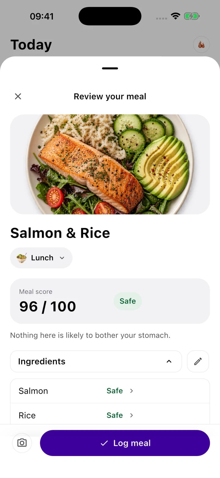
- Instant 0–100 GSI Meal Scanning: Instead of guessing whether a meal is safe, you scan your plate or log your ingredients in Gutrack to receive an instant 0–100 GSI score before you eat, complete with a breakdown of lipid load, acidity, and mechanical digestibility.
[Apple Health Biometrics]                     [Gutrack GSI Engine]
• Sleep: 5.2 hrs (Deep Sleep -35%)            • Meal Scanned: Oatmeal + Almond Butter
• Overnight RHR: +6 bpm above baseline        • Calculated Score: GSI 76 (Safe at Baseline)
• Morning HRV: 28 ms (Sympathetic Dominance)
                     │                                         │
                     └────────────────────┬────────────────────┘
                                          ▼
                       [Gutrack Clinical Correlation Insight]
   "Your mild burning today was driven by a 35% drop in deep sleep and elevated
    sympathetic tone (+6 bpm RHR), not an intolerance to oats. On high-stress days,
    buffer your meals above GSI 85 by swapping almond butter for soluble rice congee."
How Objective Data Stops Food Fear
Imagine logging a mild 4/10 burning sensation after eating your usual GSI 76 oatmeal. Without context, you panic and cross oats off your safe list.
With Gutrack, you look at your dashboard and see the objective truth: your meal scored a safe 76, which you have tolerated 14 times over the past month with zero pain. However, last night you got only 5.2 hours of sleep, your deep sleep dropped by 35%, and your overnight resting heart rate was elevated by 6 bpm.
Instantly, the psychological panic dissolves. You realize you have not developed a mysterious new food intolerance, nor is your stomach lining disintegrating. Your dorsal horn neurons are simply sensitized from sleep debt and sympathetic tone. For the next 24 hours, you adjust your target to GSI 85+ colloidal meals, prioritize vagal breathing, and return to your normal diet once your biometrics normalize.
Exporting the 14-Day Clinical PDF for Your Gastroenterologist
Gastroenterology appointments are notoriously brief—often lasting only 10 to 15 minutes. When a patient walks in with a crumpled notebook of handwritten food lists and anxious descriptions of fluctuating pain, the physician cannot easily parse the variables. The default medical response is often to double the dose of a Proton Pump Inhibitor (PPI)—which can lead to rebound acid hypersecretion (via hypergastrinemia and ECL cell hyperplasia) when you eventually try to taper off, or small intestinal bacterial overgrowth (SIBO) from chronic hypochlorhydria.
Gutrack transforms your consultation by generating a 14-Day Clinical Summary PDF designed specifically for gastroenterologists and registered dietitians. On a single, clean clinical report, your doctor can see:
- Your average daily Gastric Safety Index (GSI) adherence score and macro-distribution.
- Precise postprandial symptom onset latency (e.g., pain occurring at 15 minutes post-meal indicating mechanoreceptor stretch/visceral hypersensitivity, versus pain at 90 minutes indicating acid/pepsin accumulation or duodenal irritation).
- The direct statistical correlation between your Apple Health sleep/HRV metrics and your epigastric pain scores.
- Medication adherence (PPIs, $H_2$ blockers, alginates, or neuromodulators) mapped against symptom resolution.
Equipped with objective data showing that your symptoms correlate 85% with autonomic stress markers while your diet maintains a high GSI average, your gastroenterologist can make targeted, intelligent clinical decisions—such as avoiding unnecessary PPI escalation, evaluating for functional dyspepsia overlap, or focusing on targeted mucosal protectants (like sucralfate or zinc-L-carnosine) and vagal conditioning.
Step-by-Step Clinical Action Plan to Break the Cycle
Recovering from the intersection of gastritis and visceral hypersensitivity is not achieved through extreme starvation diets or endless anxiety over ingredients. It is achieved by systematically removing both chemical irritation and autonomic ischemia so your enteric nerves can down-regulate their firing thresholds.
- Establish a 14-Day Mucosal Baseline (GSI 75+): Use Gutrack to scan your meals and keep every meal at or above a GSI score of 75 for two consecutive weeks. Eliminate overt chemical triggers (coffee, alcohol, NSAIDs, vinegar, citrus, tomatoes, and chili peppers) so TRPV1 and ASIC3 receptors stop receiving daily proton and capsaicin bombardment.
- Adjust for High-Stress Days (GSI 85+ Protocol): Monitor your morning readiness via Apple Health inside Gutrack. On days following poor sleep ($<6.5\text{ hours}$) or acute psychological stress, shift immediately to pre-blended or gelatinized colloidal meals (bone broth congee, pureed root vegetable soups, soluble oat bran) and cap fats at $<5\text{g}$ per sitting to prevent CCK-induced stasis.
- Mandate the 5-Minute Pre-Meal Vagal Reset: Never eat in a rushed, sympathetic state. Perform 3 to 5 minutes of 4-second inhale / 6-second exhale diaphragmatic breathing before meals to unlock fundic accommodation and restore mucosal capillary blood flow.
- Eat Smaller, Volume-Controlled Meals: To avoid triggering sensitized mechanoreceptors via gastric distension, cap total meal volume (food plus liquid) at 350 mL to 400 mL per sitting, eating 4 to 5 smaller meals spaced every 3 hours rather than 2 or 3 large meals.
- Audit Your Data, Not Your Fears: When a flare occurs, resist the urge to catastrophize or permanently ban a safe food. Open Gutrack, review the meal's GSI score alongside your 48-hour sleep, RHR, and stress trendlines, and bring your 14-Day Clinical PDF to your next gastroenterology visit.
Frequently Asked Questions on the Gut-Brain Axis and Gastritis
Can stress and health anxiety cause acute gastritis flare-ups?
Yes. Acute psychological stress activates the sympathetic nervous system, which constricts splanchnic blood vessels and reduces gastric mucosal blood flow by 30% to 50%. This localized ischemia impairs epithelial cell turnover and reduces protective prostaglandin and bicarbonate secretion, leaving the stomach lining vulnerable to pepsin and hydrochloric acid degradation.
What is visceral hypersensitivity in the stomach?
Visceral hypersensitivity is a neurological state where the enteric nervous system and dorsal horn neurons become hyper-excitable following mucosal injury. In sensitized patients, normal physiological distension from 250 mL of liquid or mild pH drops ($pH\ 3.5$ to $4.5$) triggers TRPV1 pain receptors, registering in the brain
Clinical Evidence & Scientific References
- Mayer EA. Gut feelings: the emerging biology of gut-brain communication. Nat Rev Neurosci. 2011;12(8):453-466.
- Miwa H. Why dyspepsia can be painful without mucosal lesions: visceral hypersensitivity in functional dyspepsia. J Gastroenterol. 2012;47(4):352-361.
- Van Oudenhove L, et al. Biopsychosocial aspects of functional gastrointestinal disorders: How central and autonomic nervous systems drive gut sensitivity. Gastroenterology. 2016;150(6):1355-1367.
Self-tracking tool, not clinical diagnosis
Gutrack provides evidence-based Gastric Safety Index estimates for daily self-management and diet optimization. It does not replace medical consultation, endoscopy, or treatment prescribed by a qualified gastroenterologist.
Frequently asked questions
Can stress and health anxiety cause acute gastritis flare-ups?
Yes. Acute psychological stress activates the sympathetic nervous system, which constricts splanchnic blood vessels and reduces gastric mucosal blood flow by 30% to 50%. This localized ischemia impairs epithelial cell turnover and reduces protective prostaglandin and bicarbonate secretion, leaving the stomach lining vulnerable to pepsin and hydrochloric acid degradation.
What is visceral hypersensitivity in the stomach?
Visceral hypersensitivity is a neurological state where the enteric nervous system and dorsal horn neurons become hyper-excitable following mucosal injury. In sensitized patients, normal physiological distension from 250 mL of liquid or mild pH drops (pH 3.5 to 4.5) triggers TRPV1 pain receptors, registering in the brain as an 8/10 burning sensation despite normal endoscopy findings.
How does the vagus nerve influence stomach acid secretion?
The vagus nerve serves as the primary parasympathetic regulator of the upper gastrointestinal tract, driving both cephalic acid secretion via acetylcholine and protective mucosal barrier maintenance. While severe acute vagal overstimulation can transiently increase parietal cell acid output, chronic low vagal tone impairs gastric accommodation and slows antral motility, prolonging acid contact time with the mucosa.
Can tracking resting heart rate and sleep help predict gastritis flares?
Yes. Clinical tracking shows that an overnight resting heart rate (RHR) elevation of 4 to 7 bpm above baseline, or a 20% drop in heart rate variability (HRV), frequently precedes stress-induced epigastric pain by 12 to 24 hours. Correlating these autonomic markers with meal Gastric Safety Index (GSI) scores separates true food intolerances from nervous system flares.
This article is for educational purposes only and isn't a substitute for professional medical advice, diagnosis, or treatment. Gutrack is a self-tracking tool, not a medical device. If you're experiencing persistent or concerning symptoms, talk to a doctor.