Quick Answer for Erosive vs Non-Erosive Gastritis: Non-erosive gastritis is microscopic mucosal inflammation and redness (erythema) with an intact surface epithelium, typically stabilizing within 2 to 4 weeks. Erosive gastritis involves physical breaks in the superficial stomach lining above the muscularis mucosae, requiring 4 to 8 weeks of acid suppression and high-GSI (75–100) non-abrasive nutrition to achieve complete histological repair.
Not a treatment protocol
This guide has not been reviewed by a licensed clinician. Specific figures are illustrative, and no food score is a prescription. Talk to your gastroenterologist before changing your diet or medication. See our editorial standards.
When you wake up from an upper endoscopy (esophagogastroduodenoscopy, or EGD), the gastroenterologist typically hands you a preliminary printout with a few clinical photographs and a brief summary before the final pathology report arrives a week later. Two phrases appear on these reports more than almost any others: non-erosive erythematous gastritis and erosive gastritis.
To a patient experiencing daily epigastric burning, early satiety, and post-meal nausea, the distinction can feel purely academic. In fact, one of the most frustrating aspects of gastroduodenal pathology is that endoscopic severity does not always correlate linearly with symptom intensity. A patient with mild non-erosive antral erythema can experience severe visceral hypersensitivity and burning, while a patient with multiple NSAID-induced antral erosions may feel only dull aching until a lesion begins to ooze.
However, from a tissue-repair and nutritional standpoint, the difference between erosive and non-erosive disease is fundamental. It dictates how vulnerable your stomach wall is to mechanical friction, how aggressively you must buffer intragastric acidity, how your medications interact with the tissue surface, and how quickly you can safely expand your diet.
At Gutrack, we evaluate every ingredient, cooking method, and meal combination using the Gastric Safety Index (GSI)—a proprietary 0 to 100 clinical scoring algorithm that quantifies the biochemical and mechanical load of food on the upper gastrointestinal tract. By pairing your specific endoscopic diagnosis with targeted GSI thresholds, you can stop guessing why "everything feels like a trigger" and align your daily meals with the exact biological stage of your mucosal repair.
Understanding Your Endoscopy Report: Erythema vs. Erosion
To understand what your gastroenterologist saw through the endoscope—and what the pathologist observed under the microscope from your biopsy samples—it helps to review the micro-architecture of the human stomach wall.
From the inside of the stomach cavity (the lumen) moving outward, the gastric wall is organized into four distinct layers:
- The Mucosa: The innermost lining, which is further subdivided into three delicate sub-layers:
- The Surface Epithelium: A single continuous sheet of specialized columnar cells that secrete thick protective mucus and bicarbonate ions.
- The Lamina Propria: The loose connective tissue scaffolding directly beneath the epithelium that houses microscopic gastric glands (containing acid-producing parietal cells and pepsinogen-producing chief cells), delicate capillary networks, and immune cells.
- The Muscularis Mucosae: A thin ribbon of smooth muscle that forms the strict anatomical boundary between the mucosa and the deeper tissue layers.
- The Submucosa: A dense supportive layer containing larger blood vessels, lymphatics, and the Meissner nerve plexus.
- The Muscularis Propria: Thick layers of smooth muscle responsible for peristaltic contractions and mechanical grinding of food (chyme).
- The Serosa: The outer protective membrane wrapping the stomach.
Non-Erosive Gastritis: Inflammation Behind an Intact Wall
When an endoscopy report notes non-erosive gastritis (often phrased as erythematous gastropathy, patchy mucosal hyperemia, or chronic inactive/active gastritis on biopsy), the single-cell surface epithelium remains physically continuous. There are no macroscopic holes, craters, or breaks in the surface lining.
Instead, the visual redness (erythema) and swelling (edema) seen on the camera represent increased blood flow (vasodilation) and microscopic immune cell infiltration inside the underlying lamina propria.
- Active vs. Chronic on Biopsy: If the pathologist notes "chronic gastritis," they are seeing mononuclear immune cells (lymphocytes and plasma cells) in the lamina propria, indicating long-standing irritation. If they note "active gastritis," they see neutrophils invading the glandular epithelium—a hallmark of acute bacterial defense (most commonly against Helicobacter pylori) or chemical irritation.
- Clinical Vulnerability: Even though there are no open sores, the tight junctions holding the surface epithelial cells together are weakened. Hydrogen ions ($H^+$) from stomach acid can diffuse backward through these leaky junctions into the lamina propria, depolarizing sensory nerve endings and causing the classic gnawing or burning pain of gastritis.
Erosive Gastritis: Superficial Breaches in the Shield
When an endoscopy report identifies erosive gastritis (often described as multiple aphthoid erosions, hemorrhagic gastropathy, or varioliform erosions), the protective surface epithelium has physically broken down and sloughed off in localized spots.
An erosion is defined histologically as a partial-thickness mucosal defect that strips away the surface epithelium and cuts into the superficial lamina propria, without penetrating through the muscularis mucosae.
- Endoscopic Appearance: Erosions typically appear as small (usually 1 to 5 mm), flat or slightly depressed breaks in the lining. They may have a white or yellowish fibrinous base (dead epithelial cells and clotting proteins) surrounded by an angry red halo of engorged capillaries, or a dark brown/black spot (hematin) where stomach acid has oxidized a tiny pinpoint of oozing blood (subepithelial hemorrhage).
- Erosion vs. Ulcer: The muscularis mucosae is the critical anatomical dividing line. If a lesion remains above the muscularis mucosae, it is an erosion; once healed, it regenerates normal mucosal architecture without leaving a permanent fibrous scar. If the digestive process eats completely through the muscularis mucosae into the deeper submucosa (where larger arterial vessels reside), it becomes a peptic ulcer.
Pathophysiology of Erosion: How Gastric Defenses Fail
Healthy human gastric juice has a pH between 1.5 and 2.5—an environment roughly one million times more acidic than human blood (pH 7.4). Furthermore, this acid activates pepsin, a proteolytic enzyme designed to cleave collagen and dietary proteins. Why doesn't the stomach digest itself?
The answer lies in a multi-tiered defensive system known as the Gastric Mucosal Barrier:
1. The Pre-Epithelial Mucus-Bicarbonate Shield
Surface foveolar cells continuously secrete water-insoluble mucin glycoproteins (primarily MUC5AC and MUC6) that polymerize into a thick, viscoelastic gel layer adhering directly to the stomach wall. Beneath this gel, epithelial cells pump out bicarbonate ions ($HCO_3^-$).
This creates a vital micro-climate pH gradient: while the open cavity of the stomach may sit at a caustic pH of 1.8, the bicarbonate trapped inside the mucus gel neutralizes incoming hydrogen ions, keeping the immediate surface of the epithelial cells at a completely neutral pH of 6.5 to 7.0.
2. Epithelial Tight Junctions and Rapid Restitution
The surface cells are welded together near their tops by apical tight junctions (composed of claudin and occludin proteins) that block paracellular acid back-diffusion. Because these cells live in a harsh environment, the entire surface epithelium of the stomach completely renews itself every 3 to 5 days via stem cell proliferation in the neck of the gastric glands.
3. Post-Epithelial Mucosal Microcirculation and Prostaglandins
Beneath the epithelium, a dense network of capillaries in the lamina propria supplies oxygen and nutrients while carrying away any toxic acid that manages to leak through. This blood flow—along with mucus and bicarbonate secretion—is regulated continuously by local signaling molecules called prostaglandins (specifically $PGE_2$ and $PGI_2$), synthesized via cyclooxygenase (COX-1 and COX-2) enzymes.
The Cascade of Breakdown
Erosive gastritis occurs when aggressive luminal factors overwhelm or chemically disable these three defensive tiers:
- NSAIDs and Aspirin (Systemic & Topical Toxicity): Non-steroidal anti-inflammatory drugs (ibuprofen, naproxen, ketorolac, aspirin) cause erosions via a dual assault. Locally, because they are weak organic acids, they remain non-ionized in the acidic stomach lumen, allowing them to pass effortlessly through the lipid membranes of surface epithelial cells. Once inside the neutral cell interior (pH 7.0), they ionize, become trapped ("ion trapping"), and uncouple mitochondrial oxidative phosphorylation, killing the cell. Systemically, NSAIDs inhibit COX-1 enzymes, shutting down endogenous $PGE_2$ synthesis. Without prostaglandins, mucus and bicarbonate secretion plummet, and mucosal blood flow constricts, leaving the tissue ischemic and defenseless against acid.
- Alcohol and Bile Salts (Barrier Detergents): High-proof ethanol and regurgitated duodenal bile acids act as lipid detergents. They dissolve the hydrophobic phospholipids of the mucus gel layer and disrupt epithelial tight junctions, allowing massive back-diffusion of hydrogen ions into the lamina propria.
- Pepsin Activation Thresholds (pH < 4.0): Acid alone causes chemical burn, but pepsin causes structural digestion. Pepsinogen is secreted by chief cells and converts rapidly into active pepsin when intragastric pH drops below 4.0, reaching peak proteolytic violence between pH 1.5 and 2.5. Once the mucus barrier is thinned, pepsin digests the exposed cellular proteins and extracellular matrix of the lamina propria, carving out an erosion and exposing fragile subepithelial capillaries.
- Physiological Stress & Mucosal Ischemia: Severe physiological stress (major surgery, trauma, sepsis, or extreme prolonged sympathetic nervous system activation) shunts blood away from the splanchnic circulation. This microvascular ischemia starves the high-turnover gastric epithelial cells of ATP, causing rapid focal necrosis and acute stress erosions.
The 4-Stage Epithelial Healing Progression
When an erosion forms, the stomach initiates a highly coordinated biological repair sequence. Understanding these four distinct histological phases explains why your symptoms fluctuate during recovery—and why prematurely eating a heavy, acidic, or abrasive meal in Week 2 can undo ten days of cellular work in a single afternoon.
[Stage 1: Hemostasis & Gel Plug]  (Hours 0–48)
       │  Fibrin-mucoid cap seals exposed lamina propria
       ▼
[Stage 2: Acute Restitution]      (Days 2–7)
       │  Healthy neighboring cells flatten & migrate across defect
       ▼
[Stage 3: Proliferation]          (Weeks 1–3)
       │  Glandular stem cells divide to rebuild mucosal thickness
       ▼
[Stage 4: Maturation & Remodeling] (Weeks 3–8)
          Microvasculature, tight junctions & PGE2 synthesis normalize
Stage 1: Hemostasis and Formation of the Fibrin-Mucoid Cap (Hours 0 to 48)
Immediately after surface epithelial cells slough off and expose the lamina propria, damaged capillaries constrict and platelets aggregate to stop microscopic oozing. Simultaneously, necrotic cellular debris, plasma proteins (fibrinogen), and extruded mucin combine over the raw crater to form a whitish-gray fibrin-mucoid cap (often termed the "mucoid plug").
- Biological Goal: This cap acts as an emergency biological bandage. It physically shields the exposed basement membrane and fragile underlying stem cells from direct contact with luminal hydrochloric acid and pepsin.
- The Danger Zone: Active pepsin (at pH < 4.0) can enzymatically dissolve this fibrin clot, and coarse mechanical particles (such as seed hulls, raw vegetable fibers, or hard toast crusts) can physically shear the plug off the wound bed.
Stage 2: Epithelial Restitution—Cell Migration Without Division (Days 2 to 7)
Remarkably, the stomach does not wait for cell division to cover a shallow erosion. Within hours of injury—provided the luminal pH above the wound is buffered—viable epithelial cells bordering the edges of the erosion change shape. They flatten out (squamoid transformation), extend pseudopodia (lamellipodia), and migrate rapidly across the intact basement membrane underneath the fibrin-mucoid cap to re-establish a continuous single-cell sheet.
- Biological Goal: This process, called epithelial restitution, does not require mitosis (cell division); it relies solely on actin cytoskeleton rearrangement and requires an intact basement membrane and an alkaline pericellular pH maintained by underlying blood flow.
- The Danger Zone: Restitution is completely halted if intragastric acidity drops the micro-climate pH below 4.0 or if NSAIDs/alcohol impair local microvascular blood flow. Furthermore, the newly migrated cells are stretched thin and have immature tight junctions, making them acutely sensitive to high-osmolarity liquids (concentrated sugars or hypertonic broths) and capsaicin/spices.
Stage 3: Cellular Proliferation and Glandular Reconstruction (Weeks 1 to 3)
Once the surface breach is covered by the thin sheet of migrated cells, mitogenic growth factors—specifically Epidermal Growth Factor (EGF) (found abundantly in human saliva and secreted by Brunner's glands and mucosal cells) and Transforming Growth Factor-alpha (TGF-$\alpha$)—bind to receptors on progenitor stem cells located in the proliferative zone (the neck/isthmus) of adjacent gastric glands.
- Biological Goal: A surge of mitosis occurs to replace the lost cell population, restore normal columnar cell height, and rebuild the damaged upper portions of the gastric pits (foveolae).
- The Danger Zone: This is the classic "false confidence" phase for patients. Because the raw nerve endings in the lamina propria are now covered by a newly proliferated cell layer, acute burning pain often subsides by Day 10 to 14. Patients assume they are cured and eat a high-fat, acidic, or fried meal (GSI < 40). The resulting surge in cholecystokinin (CCK) delays gastric emptying for 4+ hours, bathing the thin, newly divided epithelium in prolonged acid and pepsin, stripping the nascent lining right back off.
Stage 4: Mucosal Maturation and Submucosal Remodeling (Weeks 3 to 8)
During the final phase of healing, the newly generated epithelial cells differentiate fully into specialized mucus-secreting foveolar cells. Apical tight junctions reinforce their protein cross-links, the underlying capillary network in the lamina propria remodels (angiogenesis driven by VEGF), and endogenous prostaglandin ($PGE_2$) and MUC5AC mucus synthesis return to baseline protective thickness.
- Biological Goal: Restoration of tensile strength, normal visceral nerve sensitivity thresholds, and a resilient 200-micron mucus-bicarbonate barrier capable of tolerating normal dietary variety.
Dietary Management by Healing Stage: The GSI Protocol
When patients leave an endoscopy unit with a diagnosis of erosive or non-erosive gastritis, they are routinely told to "eat a bland diet and avoid spicy, acidic, or greasy foods." This advice fails in practice because it does not quantify how foods interact with the stomach lining, nor does it account for hidden biochemical triggers in seemingly "healthy" foods.
In our clinical work at Gutrack, we see patients accidentally stall their Stage 2 and Stage 3 healing every day with foods that appear benign on paper:
- A bowl of "healthy" oatmeal topped with raw walnuts, chia seeds, and sliced strawberries (GSI 38 — High Risk due to mechanical abrasion from nuts/seeds and citric/malic acid from berries activating pepsin).
- A large dinner salad with raw kale, broccoli florets, and a lemon-olive oil vinaigrette (GSI 24 — High Risk due to insoluble cellulose shearing the fibrin-mucoid cap and pH 2.4 lemon juice immediately cleaving pepsinogen).
- A large portion of grilled ribeye steak with black pepper (GSI 31 — High Risk due to high saturated lipid content triggering CCK-mediated delayed gastric emptying, dense collagen fibers requiring hours of acid-peptic trituration, and piperine irritating TRPV1 nociceptors).
To solve this, the Gutrack Gastric Safety Index (GSI) evaluates meals across five physiological vectors on a 0 to 100 scale:
- Titratable Acidity & pH: Does the food have a pH below 4.0 that directly activates tissue-bound pepsin upon swallowing?
- Lipid Transit Delay (CCK Load): How many grams of fat (and what structure—emulsified vs. fried/saturated) are present to stimulate duodenal cholecystokinin (CCK), which slows antral peristalsis, keeps food sitting in the stomach longer, and triggers transient lower esophageal sphincter relaxations (TLESRs)?
- Mechanical & Insoluble Abrasion: Does the meal contain hard particulates, seeds, tough skins, or dense animal connective tissue that require vigorous mechanical grinding by the gastric antrum against raw erosions?
- Chemical Nociceptor Stimulation: Are there compounds like capsaicin, piperine, chlorogenic acid (in coffee), allicin (raw garlic/onion), or theobromine that trigger TRPV1 pain receptors or stimulate parietal cell gastrin release?
- Colloidal & Demulcent Protection: Does the food provide soluble beta-glucans, mucilage, or pectin that physically supplement the stomach's mucus layer?
Phase A Protocol (Days 1 to 10 of Erosive Gastritis): Target GSI 85–100
During Stages 1 and 2 (Hemostasis and Restitution), your sole nutritional objective is zero mechanical friction, rapid gastric emptying (< 90 minutes), a meal pH above 5.0, and maximal colloidal coating.
- Texture Rule: Every food must be soft enough to mash effortlessly between the tongue and the roof of the mouth, or blended into a smooth puree/soup. The gastric antrum should not have to perform mechanical grinding (trituration).
- Fat Ceiling: Strict limit of 5 to 8 grams of fat per meal, derived exclusively from gentle, non-heated sources (e.g., a half-teaspoon of extra-virgin olive oil drizzled after cooking, or the natural lipids in oatmeal and white fish).
- Volume & Temperature: Consume 5 to 6 small mini-meals (250 to 350 ml volume) rather than 3 large meals. Large meal volumes cause gastric fundic distension, which stretches the bruised mucosal wall and triggers mechanoreceptors to release gastrin, driving parietal acid output. Avoid piping hot liquids (> 55°C / 130°F), which cause thermal hyperemia on raw erosions; consume foods warm or at room temperature.
- Soluble Fiber Coating: Center meals around cooked soluble beta-glucans (rolled oats blended or cooked to a soft porridge) and cooked root starches (peeled white or sweet potatoes, carrots, kabocha squash). Beta-glucans form a viscous hydrogel in the stomach that binds excess bile salts and physically cushions the fibrin-mucoid cap.
Phase B Protocol (Days 11 to 28): Target GSI 75–85
As you enter Stage 3 (Cellular Proliferation)—or if your initial endoscopy showed non-erosive gastritis without open surface defects—you can transition from purees to intact, soft-cooked whole foods while maintaining strict protection against pepsin activation and delayed emptying.
- Protein Expansion: Introduce lean, low-collagen proteins that denature easily without prolonged acid exposure: poached egg whites (and eventually whole soft-poached eggs as lipid tolerance improves), steamed flaky white fish (cod, tilapia, halibut), skinless poached chicken breast tenders (finely shredded or minced), and silken or soft tofu.
- Carbohydrate Structure: White jasmine or basmati rice (cooked slightly soft with extra water), rice noodles, sourdough white bread (lightly toasted only to remove gummy moisture, never charred or hard), and peeled, baked apples or pears (where heat has broken down insoluble cellulose into soothing soluble pectin).
- Fat Ceiling: Gradually increase to 10 to 14 grams of gentle fat per meal (e.g., a quarter of a ripe mashed avocado, 1 teaspoon of smooth almond butter swirled into warm oats, or olive oil).
Phase C Protocol (Weeks 4 to 8+): Target GSI 65–75+
During Stage 4 (Mucosal Maturation), the goal is systematic dietary expansion without triggering a relapse.
- The One-Variable Reintroduction Rule: When testing a Caution-zone food (GSI 55–69), introduce only one new ingredient every 72 hours, always buffered inside a high-GSI base meal. Because mucosal inflammation often produces delayed symptoms (appearing 6 to 24 hours after ingestion due to cumulative acid exposure), food logging without objective meal scoring makes it nearly impossible to isolate the culprit.
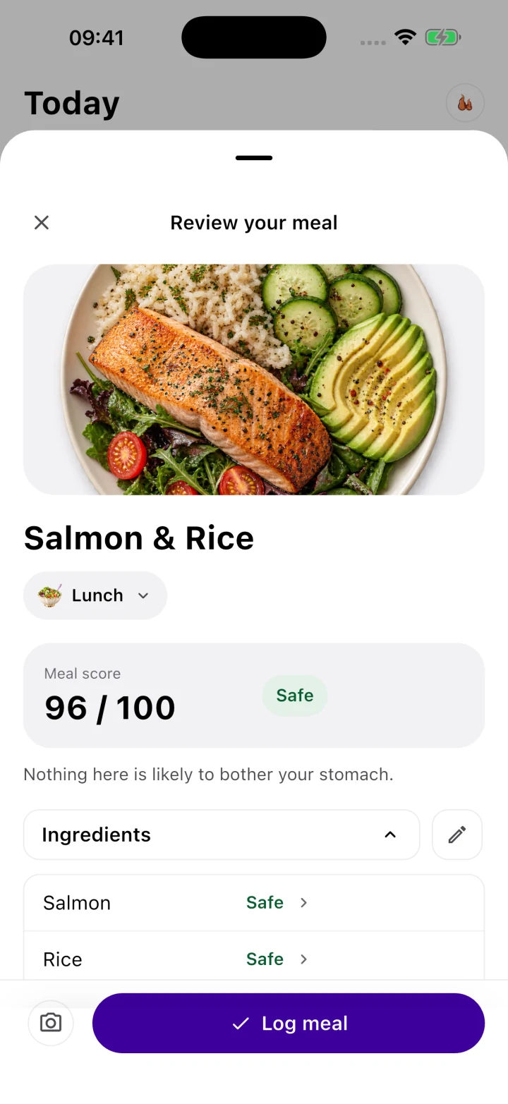
- Why Gutrack Changes Reintroduction: When you scan your plate in the Gutrack iOS app, the algorithm immediately breaks down the meal's GSI score and flags specific biochemical risks (for instance, warning you that adding garlic powder and tomato paste dropped an otherwise safe chicken-and-rice dish from a GSI of 88 down to a 42). By logging your meals alongside timestamped symptom tracking, you build an objective 14-day clinical record that reveals your exact personal GSI tolerance threshold.
GSI Reference Score Table for Erosive & Non-Erosive Healing
The table below provides clinical GSI benchmarks for specific meals and ingredients across the three safety zones, detailing the exact physiological mechanisms that help or harm a healing gastric mucosa.
| Meal / Food Item | GSI Score (0–100) | Safety Zone | Healing Stage Suitability | Biochemical & Physiological Mechanism |
|---|---|---|---|---|
| Warm Rolled Oat Porridge with Marshmallow Root Infusion & Mashed Banana (made with water or fortified oat milk) | 96 / 100 | Safe (Optimal) | Stages 1–4 (Acute Erosive & Non-Erosive) | High concentration of soluble beta-glucans and mucilage polysaccharides forms a viscoelastic colloidal hydrogel over exposed lamina propria; pH ~6.2 buffers luminal acid; low lipid load (< 3g) ensures rapid antral emptying without CCK delay. |
| Steamed Cod Fillet over Pureed Butternut Squash & Soft White Jasmine Rice | 92 / 100 | Safe (Optimal) | Stages 1–4 (Late Stage 1 onward) | Flaky white fish has minimal stromal elastin/collagen, requiring minimal pepsin proteolysis; cooked squash provides gentle soluble pectin and beta-carotene (supporting epithelial turnover); zero insoluble mechanical abrasion. |
| Poached Chicken Breast (Shredded) in Homemade Bone/Vegetable Broth with Rice Noodles & Peeled Zucchini | 88 / 100 | Safe | Stages 2–4 | Warm isotonic liquid broth hydrates mucosa; amino acids (particularly L-glutamine and glycine) provide direct metabolic fuel for proliferating glandular neck cells; peeled zucchini eliminates insoluble skin friction. |
| Baked Peeled Apple with a Pinch of Ceylon Cinnamon & Silken Tofu Custard | 84 / 100 | Safe | Stages 2–4 | Thermal baking converts rigid apple protopectin into soothing soluble pectin and softens malic acid sharpness; silken tofu offers high-bioavailability plant protein with an alkaline buffering profile and zero mechanical grit. |
| Soft-Poached Whole Egg on Lightly Toasted White Sourdough with 1/4 Mashed Avocado | 76 / 100 | Safe (Transition) | Stages 3–4 (and Stable Non-Erosive) | Sourdough fermentation degrades fructans and phytic acid, improving gastric digestibility; egg yolk and avocado introduce moderate emulsified monounsaturated fats (~11g total), gently testing CCK transit without overwhelming the LES. |
| Plain Greek Yogurt (2% Fat) with Raw Blueberries & Chopped Walnuts | 56 / 100 | Caution | Stage 4 Only (Test Cautiously) | Lactic acid lowers yogurt pH to ~4.3; blueberry skins and citric acid (pH ~3.2) can reactivate surface-bound pepsin; rigid walnut particulates require vigorous antral grinding that can abrade immature Stage 2/3 epithelium. |
| Pan-Seared Salmon with Quinoa & Roasted Asparagus Spears | 52 / 100 | Caution | Stage 4 Only | Although rich in anti-inflammatory omega-3s, a thick salmon fillet delivers 18–22g of lipid, slowing gastric emptying via CCK; quinoa saponin coatings and fibrous asparagus stalks create moderate mechanical drag across the antrum. |
| Decaf Black Coffee or Green Tea on an Empty Stomach | 34 / 100 | High Risk | Avoid in Stages 1–3 | Even without caffeine, coffee contains chlorogenic acids and roasting compounds (N-alkanoyl-5-hydroxytryptamides) that potently stimulate parietal cell gastrin and hydrochloric acid secretion upon contact with an empty, unbuffered mucosa. |
| Raw Kale Salad with Cherry Tomatoes, Red Onion & Lemon Vinaigrette | 22 / 100 | High Risk | Avoid during Active Healing | Lemon juice (pH 2.2) and tomatoes (pH 4.1) immediately trigger maximal pepsinogen cleavage; tough insoluble cellulose in raw kale physically shears the fibrin-mucoid cap; raw onion disulfides activate TRPV1 pain fibers. |
| Spicy Pepperoni Pizza or Fried Chicken with Garlic Dipping Sauce & Soda | 11 / 100 | High Risk (Severe) | Strictly Contraindicated | High saturated fat (> 30g) induces prolonged gastric stasis (4–6 hours) and TLESRs; capsaicin and allicin inflame exposed lamina propria capillaries; carbonated phosphoric/citric acid distends the stomach and drops pH < 2.5. |
Medication Timing, Cytoprotection, and Daily Tracking
In erosive gastritis, nutrition and pharmacology work as partners. Medication temporarily alters the intragastric chemical environment or creates a synthetic physical barrier, giving your epithelial stem cells the quiet biological window they need to execute Stages 1 through 4 of repair. However, improper medication timing—or pairing medications with low-GSI meals—routinely blunts their effectiveness.
1. Proton Pump Inhibitors (PPIs): Omeprazole, Pantoprazole, Esomeprazole
PPIs are prodrugs that are absorbed in the small intestine, travel through the bloodstream, and accumulate in the secretory canaliculi of active parietal cells, where they irreversibly bind to and shut down the $H^+/K^+$ ATPase enzyme (the proton pump).
- The pH > 4.0 Clinical Target: For an erosion to heal rapidly, intragastric pH must be held above 4.0 for at least 16 to 18 hours a day. This keeps pepsin inactive and prevents acid from dissolving the fibrin-mucoid cap.
- Timing Rule: PPIs only deactivate proton pumps that are actively inserted into the cell membrane waiting for food. Therefore, a PPI must be taken 30 to 60 minutes before your first true meal of the day (typically breakfast). Taking a PPI at bedtime or after a meal misses peak pump activation, cutting its acid-suppressing efficacy by up to 40%.
- Navigating Rebound Acid Hypersecretion (RAHS): When you take a PPI for more than 4 to 8 weeks, your stomach compensates for the lack of acid by secreting extra gastrin, which causes parietal and enterochromaffin-like (ECL) cells to hypertrophy (grow larger and more numerous). If you stop your PPI abruptly once your erosions heal, those enlarged cell populations unleash a massive surge of rebound acid 5 to 14 days later, often causing new erosions. Always work with your physician on a structured step-down taper (e.g., transitioning to every-other-day dosing or bridging with an H2 blocker like famotidine) while keeping your diet strictly in the GSI 80+ Safe Zone during the taper window.
2. Sucralfate (Carafate): The Prescription Liquid Bandage
For erosive gastritis and bile reflux gastropathy, sucralfate (an aluminum hydroxide complex of sulfated sucrose) is one of the most targeted mucosal cytoprotectants available.
- Mechanism of Action: When sucralfate enters an acidic environment, it polymerizes into a thick, sticky, viscid paste. Critically, this paste carries a strong negative electrical charge. Because the exposed proteins (albumin and fibrinogen) inside the raw crater of a gastric erosion carry a positive electrical charge, sucralfate binds electrostatically and selectively directly onto the open erosion for up to 6 hours—shielding it from acid, pepsin, and bile salts while stimulating local prostaglandin ($PGE_2$) and epidermal growth factor (EGF) release.
- Timing Rule: Sucralfate must be taken on an empty stomach—specifically 1 hour before meals and at bedtime. If you take sucralfate with food, the positively charged proteins in your meal bind up the medication in the lumen before it can ever reach the stomach wall. Furthermore, separate sucralfate by at least 30 to 60 minutes from PPIs or antacids.
3. H. pylori Eradication Therapy Considerations
If your biopsy reveals that Helicobacter pylori is driving your erosive or non-erosive gastritis, your gastroenterologist will prescribe a 10-to-14-day regimen of high-dose antibiotics alongside a PPI and often bismuth subsalicylate (quadruple therapy).
- Bismuth acts locally to coat erosions and disrupt bacterial cell walls (Note: bismuth harmlessly turns stools dark black, which should be distinguished from true melena).
- Because broad-spectrum antibiotics can irritate the stomach lining and alter gut motility, maintaining a GSI 85+ diet throughout the entire 14-day antibiotic course is essential to prevent treatment-limiting nausea and epigastric cramping.
Why Traditional Food Diaries Fail—and How to Track Objectively
If you have ever tried keeping a paper notebook or a basic calorie-tracking food log during a gastritis flare, you already know the frustration: everything starts to look like a trigger.
You eat a gentle breakfast at 8:00 AM, a questionable lunch at 1:00 PM, and a completely safe dinner at 7:00 PM—yet you wake up at 2:00 AM with severe epigastric burning. Most patients blame the safe 7:00 PM dinner, cross those safe foods off their list, and progressively shrink their diet down to three or four foods, sliding toward nutritional deficiency and severe food anxiety.
What actually happened physiologically? The 1:00 PM lunch (perhaps containing hidden acidity, garlic powder, or excessive fat) stripped away the fragile Stage 2 epithelial layer or thinned the mucus barrier. Once that barrier was compromised, the normal circadian surge of nocturnal histamine-driven acid secretion between 11:00 PM and 2:00 AM burned the newly exposed lamina propria nerves. The 7:00 PM dinner was innocent—it simply landed on tissue that had been sensitized six hours earlier.
The Gutrack Clinical Advantage
We built Gutrack (available on iOS at gutrack.com) specifically to eliminate this guesswork for patients with erosive gastritis, non-erosive gastritis, and GERD:
- Instant 0–100 GSI Meal Scanning: Before you take a bite, scan or log your meal in Gutrack. The app calculates the exact Gastric Safety Index (0–100) score, evaluating acidity, fat transit load, mechanical texture, and chemical irritants so you know immediately whether a meal fits your current healing stage.
- Delayed-Trigger Timeline Correlation: Log symptoms (epigastric burning, early satiety, nausea, reflux, bloating) alongside medication doses (PPIs, sucralfate, H2 blockers). Gutrack correlates symptom spikes not just with your last meal, but with cumulative GSI load over the preceding 24 to 48 hours.
- 14-Day Clinical PDF Exports for Your Gastroenterologist: Instead of walking into your 15-minute gastroenterology follow-up trying to summarize a messy notebook, export a clean, structured 14-Day Gutrack Clinical Report. Your physician can instantly view your average daily GSI score, medication adherence, symptom frequency curve, and top biochemical triggers—allowing them to make evidence-based decisions on PPI tapering or repeat endoscopy without guessing.
Clinical Summary Checklist: Erosive vs. Non-Erosive Recovery
To ensure your mucosal lining progresses smoothly from acute injury back to full structural resilience, follow these core clinical benchmarks:
| Clinical Parameter | Non-Erosive Gastritis Protocol | Erosive Gastritis Protocol |
|---|---|---|
| Histological State | Intact surface epithelium; lamina propria immune cell infiltration & erythema. | Focal physical breaks in surface epithelium & superficial lamina propria (above muscularis mucosae). |
| Expected Healing Window | 2 to 4 weeks with trigger |
Self-tracking tool, not clinical diagnosis
Gutrack provides evidence-based Gastric Safety Index estimates for daily self-management and diet optimization. It does not replace medical consultation, endoscopy, or treatment prescribed by a qualified gastroenterologist.
Frequently asked questions
What is the clinical difference between erosive and non-erosive gastritis?
Non-erosive gastritis involves intact surface tissue showing microscopic white blood cell infiltration and endoscopic redness (erythema), most commonly driven by Helicobacter pylori or mild bile reflux. Erosive gastritis features visible, shallow physical breaks in the superficial mucosal layer (Typically < 3 to 5 mm in depth) that do not penetrate the underlying muscularis mucosae layer.
Can erosive gastritis turn into an ulcer?
Yes, if the offending trigger (such as NSAIDs, alcohol, or unmanaged acid-peptic exposure) continues, superficial erosions can digest through the muscularis mucosae into the deeper submucosa. Once a lesion exceeds 5 mm in diameter and penetrates past the muscularis mucosae, it is clinically reclassified as a frank peptic ulcer, which carries higher risks of scarring and bleeding.
How long does erosive gastritis take to heal on endoscopy?
Superficial epithelial restitution begins within 24 to 48 hours of removing the chemical trigger, with acute mucosal defects closing within 7 to 14 days under optimal pH control (pH > 4.0). However, complete submucosal architectural remodeling and restoration of normal prostaglandin and mucus-bicarbonate secretion typically require 4 to 8 weeks.
What are the warning signs of gastric bleeding with erosive gastritis?
Because erosive gastritis exposes the superficial capillary network within the lamina propria, multiple erosions can cause slow or acute oozing. Warning signs requiring immediate emergency evaluation include melena (black, tarry, foul-smelling stools), hematemesis (vomiting bright red blood or dark 'coffee-ground' material), unexplained dizziness upon standing, or sudden shortness of breath.
Sources & background reading
Background source for definitions, diagnosis and treatment. Food scores are estimates, not clinical targets. See our editorial standards.
Clinical Evidence & Scientific References
- Rugge M, et al. OLGA staging for gastritis: a tutorial. Dig Liver Dis. 2008;40(8):650-658.
- Laine L, et al. Stratifying risk and managing mucosal erosions in acute gastric injury. Gastroenterology. 2008;135(1):41-60.
- American Gastroenterological Association (AGA). Institute Guideline on the Management of Gastric Subepithelial Lesions and Erosions. 2022.
This article is for educational purposes only and isn't a substitute for professional medical advice, diagnosis, or treatment. Gutrack is a self-tracking tool, not a medical device. If you're experiencing persistent or concerning symptoms, talk to a doctor.